Copyright
©2005 Baishideng Publishing Group Co.
World J Gastroenterol. Jan 14, 2005; 11(2): 216-220
Published online Jan 14, 2005. doi: 10.3748/wjg.v11.i2.216
Published online Jan 14, 2005. doi: 10.3748/wjg.v11.i2.216
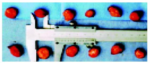
Figure 1 Tumors in nude mice on the 30th d.
Upper line of tumors was HCC in the control group, the lower in therapy group.
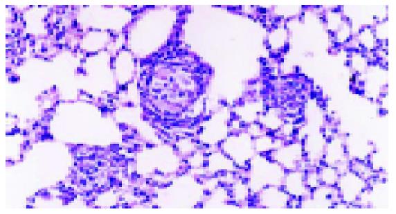
Figure 2 Metastases of hepatocellular carcinoma in lungs.
Magnification: ×200.
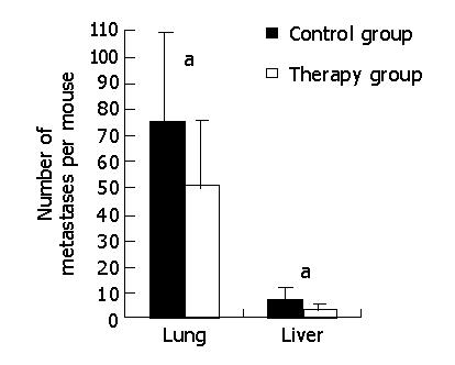
Figure 3 Metastases of hepatocellular carcinoma in lungs and liver.
aP<0.05. Liver metastases were recorded in a gross manner by examining each lobe of liver and counting macroscopic tumors on the surface. Lung metastases were counted under microscope by observing consecutive paraffin slices of lung.
Figure 4 Immunohistochemical expression of CD34.
A: control group; B: thalidomide treated group. Magnification: ×200.
Figure 5 RT-PCR of VEGF mRNA in HCC tissue.
M: Marker, 100 bp DNA ladder, ranging from 100 bp to 600 bp; C: Control group; T: Therapy group. The band of VEGF (all isoforms, 196 bp) and G3PDH (450 bp) are shown at expected location in the gel. G3PDH used as an internal standard. VEGF mRNA was expressed strongly in control group, whereas weakly in therapy group.
- Citation: Zhang ZL, Liu ZS, Sun Q. Effects of thalidomide on angiogenesis and tumor growth and metastasis of human hepatocellular carcinoma in nude mice. World J Gastroenterol 2005; 11(2): 216-220
- URL: https://www.wjgnet.com/1007-9327/full/v11/i2/216.htm
- DOI: https://dx.doi.org/10.3748/wjg.v11.i2.216


