Copyright
©2005 Baishideng Publishing Group Inc.
World J Gastroenterol. Apr 28, 2005; 11(16): 2444-2449
Published online Apr 28, 2005. doi: 10.3748/wjg.v11.i16.2444
Published online Apr 28, 2005. doi: 10.3748/wjg.v11.i16.2444
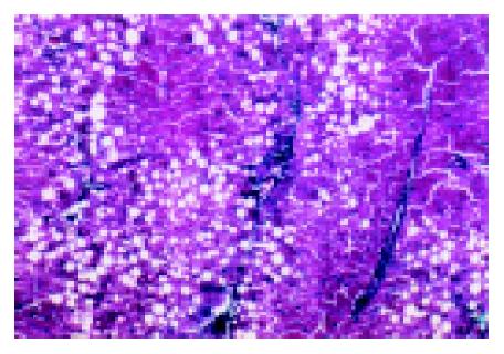
Figure 1 Liver fibrosis with inflammation necrosis and ballooning after CCl4 injection for 9 wk (Masson, 100×).
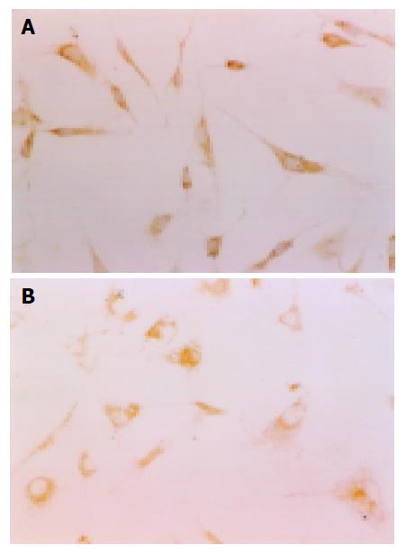
Figure 2 HSCs in the media of different sera (immunocytochemistry stain of α-SMA, 400×).
A: HSCs in NCS/RPMI-1640, B: HSCs in drug sera/RPMI-1640: cells expanded and synapses became fewer and smaller.
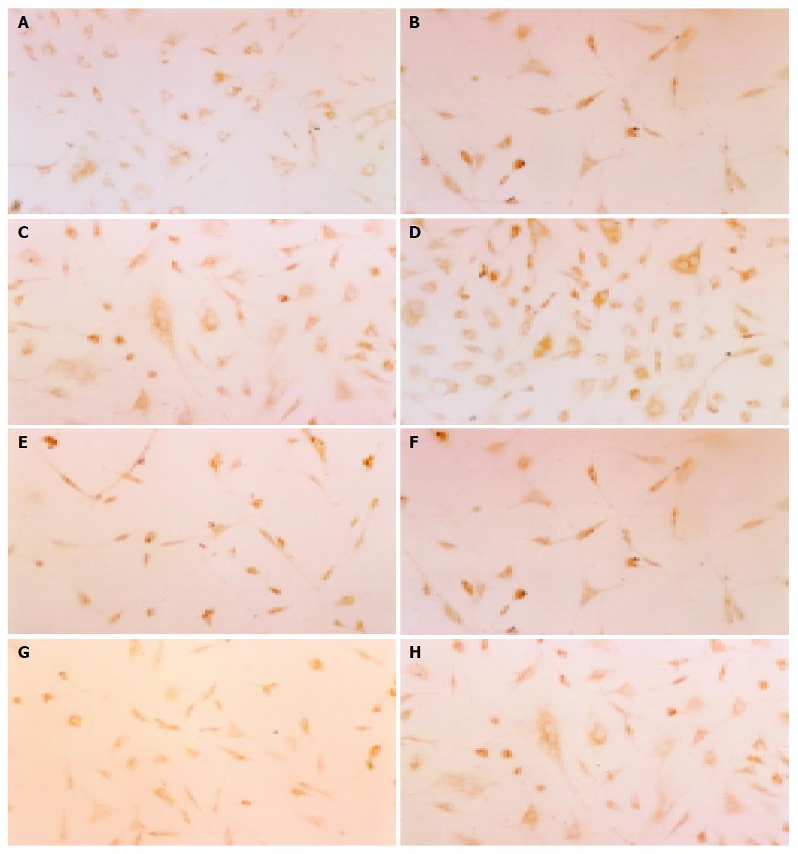
Figure 3 Immunocytochemistry stain of α-SMA in HSCs-positive cells: brown in plasm.
When HSCs were inhibited, the number of HSCs decreased, while the expression of α-SMA increased, with cell bodies expanded and synapses fewer and smaller (200×). A: HSCs in normal serum of Salvia miltiorrhiza; B: HSCs in normal serum of Yigankang; C: HSCs in normal serum of colchicines; D: HSCs in normal serum of rats; E: HSCs in pathological serum of Salvia miltiorrhiza; F: HSCs in pathological serum of Yigankang; G: HSCs in pathological serum of colchicines; H: HSCs in pathological serum of fibrotic rats.
- Citation: Yao XX, Lv T. Effects of pharmacological serum from normal and liver fibrotic rats on HSCs. World J Gastroenterol 2005; 11(16): 2444-2449
- URL: https://www.wjgnet.com/1007-9327/full/v11/i16/2444.htm
- DOI: https://dx.doi.org/10.3748/wjg.v11.i16.2444


