Copyright
©2005 Baishideng Publishing Group Inc.
World J Gastroenterol. Apr 28, 2005; 11(16): 2413-2419
Published online Apr 28, 2005. doi: 10.3748/wjg.v11.i16.2413
Published online Apr 28, 2005. doi: 10.3748/wjg.v11.i16.2413
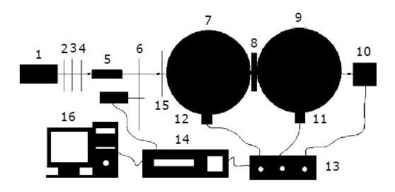
Figure 1 Experimental set-up consisting of double- integrating-sphere with an intervening sample.
1. Laser 2. Attenuator 3. Attenuator 4. 2 mm pinhole 5. Beam expander 6. 6 mm pinhole 7. Integrating sphere 8. Sample 9. Integrating sphere 10. Photodiode detector 11. Photodiode detector 12. Photodiode detector 13. Switch box 14. Lock-in-amplifier 15. Light chopper 16. PC.
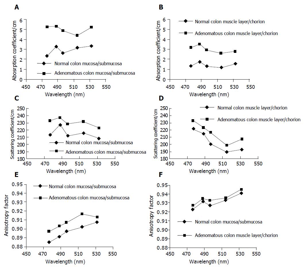
Figure 2 Optical properties of normal and adenomatous human colon mucosa/submucosa in vitro vary with a change of laser wavelength in vitro.
A: Plots of the absorption coefficients measured in vitro vs wavelength of normal and adenomatous human colon mucosa/submucosa; B: Plots of the absorption coefficients measured in vitro vs wavelength of normal and adenomatous human colon muscle layer/chorion; C: Plots of the scattering coefficients measured in vitro vs wavelength of normal and adenomatous human colon mucosa/submucosa; D: Plots of the scattering coefficients measured in vitro vs wavelength of normal and adenomatous human colon muscle layer/chorion; E: Plots of the anisotropy factors measured in vitro vs wavelength of normal and adenomatous human colon mucosa/submucosa; F: Plots of the anisotropy factors measured in vitro vs wavelength of normal and adenomatous human colon muscle layer/chorion.
-
Citation: Wei HJ, Xing D, Lu JJ, Gu HM, Wu GY, Jin Y. Determination of optical properties of normal and adenomatous human colon tissues
in vitro using integrating sphere techniques. World J Gastroenterol 2005; 11(16): 2413-2419 - URL: https://www.wjgnet.com/1007-9327/full/v11/i16/2413.htm
- DOI: https://dx.doi.org/10.3748/wjg.v11.i16.2413


