Copyright
©2005 Baishideng Publishing Group Inc.
World J Gastroenterol. Apr 21, 2005; 11(15): 2364-2366
Published online Apr 21, 2005. doi: 10.3748/wjg.v11.i15.2364
Published online Apr 21, 2005. doi: 10.3748/wjg.v11.i15.2364
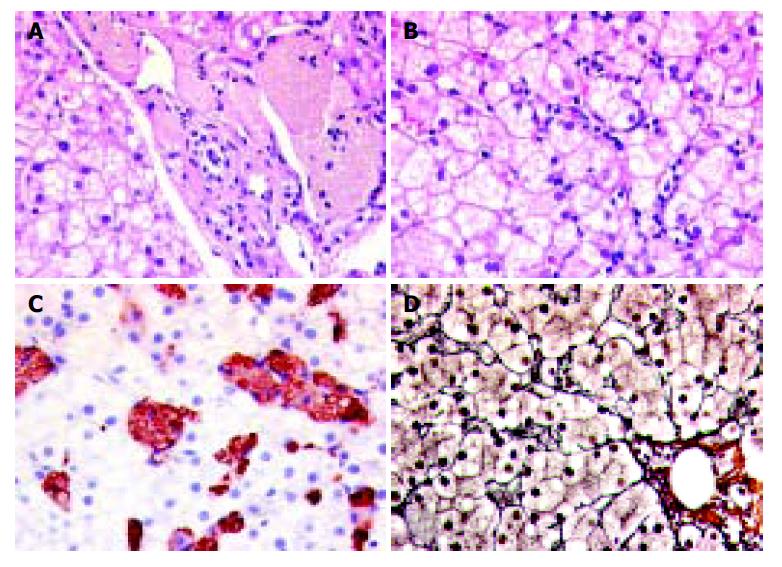
Figure 1 Light microscopic findings in the liver.
A: A tan-colored foamy material is seen in conspicuous macrophages next to and in a portal tract. HE stain ×400. B: Part of an acinus with a prominent intrasinusoidal cellular infiltration and a microvesicular steatosis of hepatocytes. HE stain (×400). C: An immunohistochemical staining with CD68 positive macrophages laden with the foamy material. D: Reticulin stain (×400). No remarkable fibrosis.
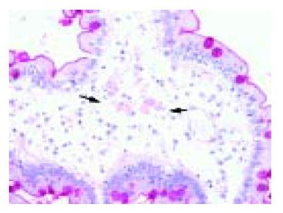
Figure 2 Small bowel biopsy reveals single macrophages with cytoplasmatic storage of PAS-positive foamy, tan-colored material within the apical part of the villi in the lamina submucosa.
A PAS stain is demonstrated with PAS-positive macrophages between the arrows (cd) (×400).
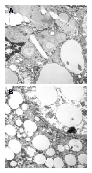
Figure 3 A: Electron micrograph of liver tissue demonstrates a single cholesterol crystal in the cytoplasm of a liver cell.
B: Triglyceride droplets of varied size in the cytoplasm of hepatocytes (×11000).
- Citation: Drebber U, Andersen M, Kasper HU, Lohse P, Stolte M, Dienes HP. Severe chronic diarrhea and weight loss in cholesteryl ester storage disease: A case report. World J Gastroenterol 2005; 11(15): 2364-2366
- URL: https://www.wjgnet.com/1007-9327/full/v11/i15/2364.htm
- DOI: https://dx.doi.org/10.3748/wjg.v11.i15.2364


