Copyright
©2005 Baishideng Publishing Group Inc.
World J Gastroenterol. Apr 14, 2005; 11(14): 2088-2094
Published online Apr 14, 2005. doi: 10.3748/wjg.v11.i14.2088
Published online Apr 14, 2005. doi: 10.3748/wjg.v11.i14.2088
Figure 1 Expression, purification, and Western blot analysis of eight GST fusion proteins containing overlapping preS1 fragments in E.
coli BL21 (gold). A and B: Cell lysates and purified fusion proteins subjected to 12.5% SDS-PAGE and stained with Coomassie brilliant blue. C: Protein bands on the duplicate gel of cell lysates electroblotted onto a nitrocellulose membrane for Western blot analysis with mice antisera. Lane M: Molecular weight standard. Lanes 1-8: The samples containing preS1 (21-119), preS1 (21-47), preS1 (34-59), preS1 (48-70), preS1 (60-85), preS1 (71-94), preS1 (86-109) and preS1 (95-119) in a sequent order.
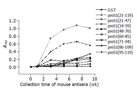
Figure 2 Immunogenicity analysis of different fragments in preS1 (21-119) region in mice by ELISA using eight overlapping GST-preS1 fusion proteins as coated antigens.
The mice were immunized with His-preS1 (21-119) fusion proteins at wk 0, 2, 4, 6 and 8, and the antisera were collected the following week.
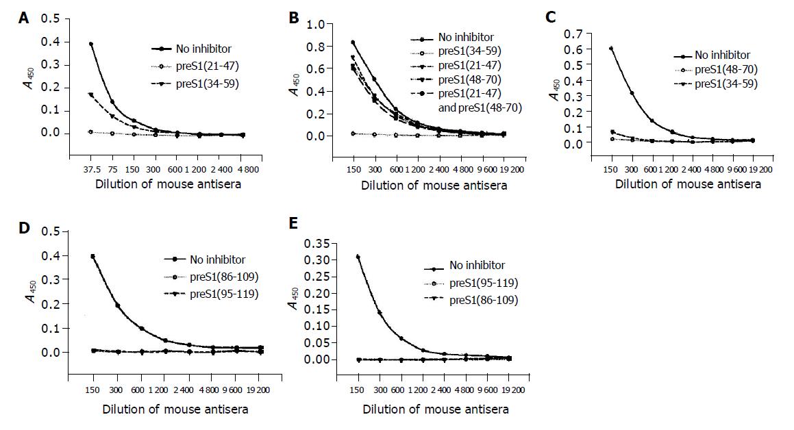
Figure 3 Identification of the precise immunogenic domains in mice by competitive ELISA.
The coated antigens were the five fusion proteins containing different preS1 fragments: preS1 (21-47) (A), preS1 (34-59) (B), preS1 (48-70) (C), preS1 (86-109) (D) and preS1 (95-119) (E), respectively. The mouse antisera colleted at wk 9 were previously incubated with the coated antigens or the other overlapping antigen(s) at a dose of 2 µg, and the samples not treated with antigen(s) were used as controls.
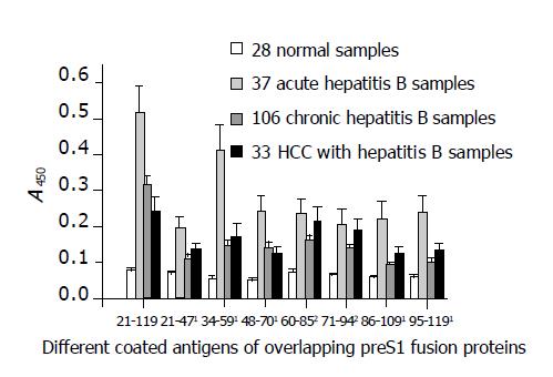
Figure 4 Detection of antigenicity (A450) of the eight different preS1 fragments overlapped in preS1 (21-119) region against hepatitis B patients’ serum samples with different infection types by indirect ELISA.
1The antigenicity of the fragments, preS1 (21-47), preS1 (48-70), preS1 (86-109), preS1 (95-119), and especially preS1 (34-59), decreased significantly in chronic-phase sera (chronic hepatitis B and HCC with hepatitis B samples) in comparison with acute-phase sera (P<0.05). However, 2the antigenicity of the fragments, preS1 (60-85) and preS1 (71-94), in the middle part showed less diversity (P>0.05). Each column represents the mean A value±SE.
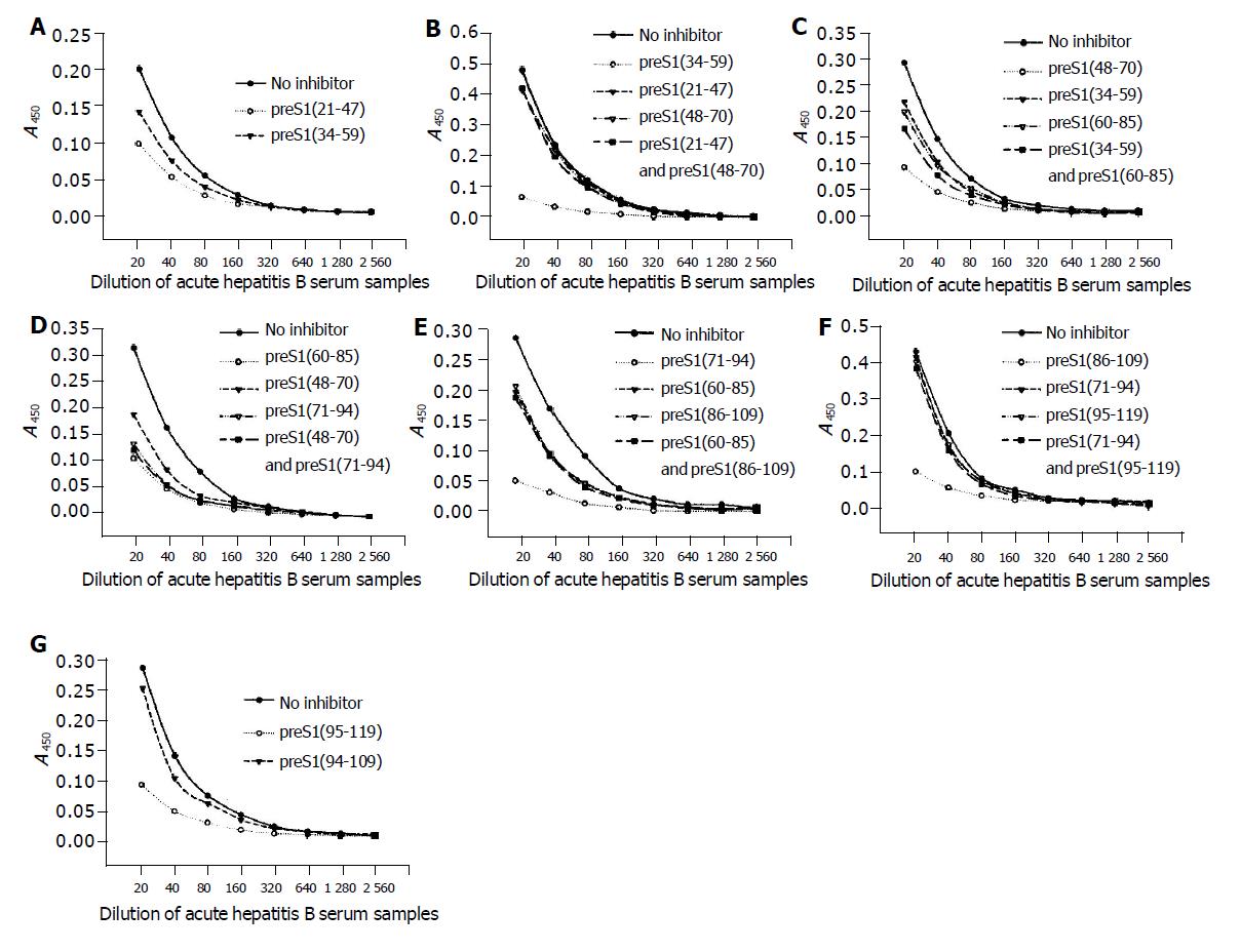
Figure 5 Identification of the precise immunogenic domains in humans by competitive ELISA.
The coated antigens were the seven fusion proteins containing different preS1 fragments: preS1 (21-47) (A), preS1 (34-59) (B), preS1 (48-70) (C), preS1 (60-85) (D), preS1 (71-94) (E), preS1 (86-109) (F) and preS1 (95-119) (G), respectively. The mixture of sera samples from acute hepatitis B patients was previously incubated with the coated antigens or the overlapping antigen(s) at a dose of 2 µg, and the samples not treated with antigen(s) were used as controls.
- Citation: Hu WG, Wei J, Xia HC, Yang XX, Li F, Li GD, Wang Y, Zhang ZC. Identification of the immunogenic domains in HBsAg preS1 region using overlapping preS1 fragment fusion proteins. World J Gastroenterol 2005; 11(14): 2088-2094
- URL: https://www.wjgnet.com/1007-9327/full/v11/i14/2088.htm
- DOI: https://dx.doi.org/10.3748/wjg.v11.i14.2088


