Copyright
©2005 Baishideng Publishing Group Inc.
World J Gastroenterol. Apr 7, 2005; 11(13): 1946-1950
Published online Apr 7, 2005. doi: 10.3748/wjg.v11.i13.1946
Published online Apr 7, 2005. doi: 10.3748/wjg.v11.i13.1946
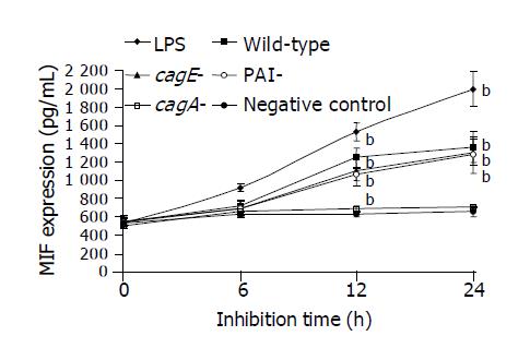
Figure 1 Expression of MIF by THP1 after co-culture with LPS (1 ng/mL) and whole cell proteins (40 μg/mL) of wild-type cytotoxic H pylori strain TN2 and its isogenic mutants, TN2△cag (PAI-), TN2△cagA (cagA-) and TN2△cagE (cagE-).
The bars represent the standard deviations. bP<0.01, compared with the control.
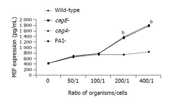
Figure 2 Expression of MIF by THP1 after co-culture with a wild-type cytotoxic H pylori strain TN2 and its isogenic mutants, TN2△cag (PAI-), TN2△cagA (cagA-) and TN2△cagE (cagE-), at different organism/cell ratios for 12 h.
The bars represent the standard deviations. bP<0.01, compared with the isogenic mutant TN2△cag (PAI-).
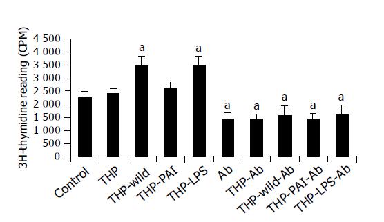
Figure 3 Effect of LPS and H pylori-co-cultured supernatants of THP1 cells, with and without addition of monoclonal anti-MIF antibody (Ab), on the cell proliferation of MKN-45.
The bars represent the standard deviations. aP<0.05 vs the control.
-
Citation: Xia HHX, Lam SK, Chan AO, Lin MCM, Kung HF, Ogura K, Berg DE, Wong BCY. Macrophage migration inhibitory factor stimulated by
Helicobacter pylori increases proliferation of gastric epithelial cells. World J Gastroenterol 2005; 11(13): 1946-1950 - URL: https://www.wjgnet.com/1007-9327/full/v11/i13/1946.htm
- DOI: https://dx.doi.org/10.3748/wjg.v11.i13.1946


