Copyright
©2005 Baishideng Publishing Group Inc.
World J Gastroenterol. Jan 7, 2005; 11(1): 84-88
Published online Jan 7, 2005. doi: 10.3748/wjg.v11.i1.84
Published online Jan 7, 2005. doi: 10.3748/wjg.v11.i1.84
Figure 1 Expression of TGIF protein by cell immunohistochemistry.
A: cells stably transfected with pcDNA3.1-TGIF; B: cells stably transfected with PcDNA3.1; C: parental cells.
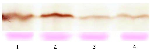
Figure 2 Expression of TGIF protein by Western blotting.
Lanes 1 and 2: TGIF expressing cells; lane 3: vector control cells, lane 4: parental cells. The lower panel was stained with Ponceau S as a loading control.
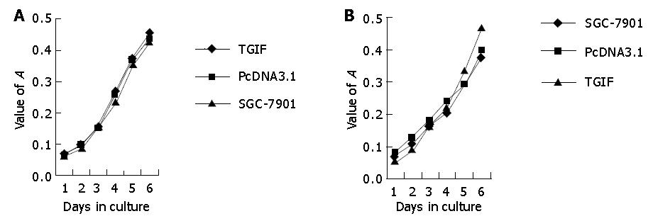
Figure 3 Proliferation rate of TGIF transfected, vector control and parental cells.
A: Without 10 μg/L TGF-1; B: with 10 μg/L TGF-β 1.
Figure 4 Plating efficiency in parental, vector control and TGIF transfected cells.
Figure 5 Morphology of blank, negative control and TGIF transfectant cells by TEM×15000.
A: parental cell; B: vector control cell; C: TGIF transfectant cell.
Figure 6 Tumor development in nude mice.
A: mice inoculated with parental cells; B: mice inoculated with vector control cells; C: mice inoculated with TGIF expressing cells.
- Citation: Hu ZL, Wen JF, Xiao DS, Zhen H, Fu CY. Effects of transforming growth interacting factor on biological behaviors of gastric carcinoma cells. World J Gastroenterol 2005; 11(1): 84-88
- URL: https://www.wjgnet.com/1007-9327/full/v11/i1/84.htm
- DOI: https://dx.doi.org/10.3748/wjg.v11.i1.84


