Copyright
©2005 Baishideng Publishing Group Inc.
World J Gastroenterol. Jan 7, 2005; 11(1): 7-16
Published online Jan 7, 2005. doi: 10.3748/wjg.v11.i1.7
Published online Jan 7, 2005. doi: 10.3748/wjg.v11.i1.7
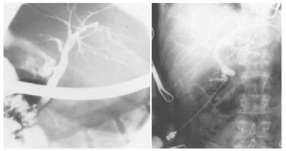
Figure 1 Cholangiographic pictures of enlarged bile ducts in a PSC patient.
On the left picture of ERCP, and on the right picture of PTC, multifocal stricturing and slightly dilated bile ducts are visible in both pictures.
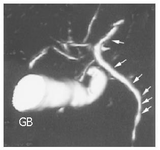
Figure 2 MRCP pictures of a PSC patient.
Wall irregularities (see arrows) are visible in undilated bile ducts. The gallbladder (GB) is enlarged.
Figure 3 Histological appearance of the common bile duct (A) and a large intralobular bile duct (B) in PSC (Cross section of liver, 4× and 40× magnification, Masson Stain).
Figure 4 Histological appearance of a small bile duct with inflammatory cells (A) and a small intra-hepatic bile duct with concentric rings of fibrosis (B) in PSC (40× magnification, H&E).
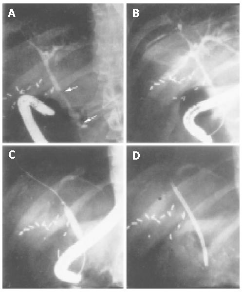
Figure 5 Sequence of balloon dilatation during ERCP treatment in a PSC patient with prior multiple bile duct strictures.
- Citation: Portincasa P, Vacca M, Moschetta A, Petruzzelli M, Palasciano G, van Erpecum KJ, van Berge-Henegouwen GP. Primary sclerosing cholangitis: Updates in diagnosis and therapy. World J Gastroenterol 2005; 11(1): 7-16
- URL: https://www.wjgnet.com/1007-9327/full/v11/i1/7.htm
- DOI: https://dx.doi.org/10.3748/wjg.v11.i1.7


