Copyright
©The Author(s) 2004.
World J Gastroenterol. May 1, 2004; 10(9): 1361-1364
Published online May 1, 2004. doi: 10.3748/wjg.v10.i9.1361
Published online May 1, 2004. doi: 10.3748/wjg.v10.i9.1361
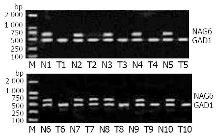
Figure 1 Expression of NAG6 in gastric carcinoma and corre-sponding normal tissues examined by RT-PCR.
The RT prod-ucts were examined by PCR with NAG6 primers, producing a 680 bp fragment and with GAPDH primers, producing a 466 bp fragment. Lane M: 2 000 bp marker, Lane N: normal epithe-lium tissues, Lane T: gastric carcinoma tissues.
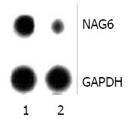
Figure 2 Dot hybridization analysis of NAG6 gene expression profiles in human gastric carcinoma and corresponding nor-mal tissues.
NAG6 cDNA obtained by RT-PCR was blotted onto nylon membranes. The membranes were hybridized with 32P-labeled cDNA probes obtained from total RNA of human gas-tric carcinoma (1) and corresponding normal gastric epithelial; (2) tissues. After stringent washes, membranes were exposed to X-ray film for 4 d at-70 °C. NAG6 was down-regulated in gastric carcinoma tissues.
- Citation: Zhang XM, Sheng SR, Wang XY, Bin LH, Wang JR, Li GY. Expression of tumor related gene NAG6 in gastric cancer and restriction fragment length polymorphism analysis. World J Gastroenterol 2004; 10(9): 1361-1364
- URL: https://www.wjgnet.com/1007-9327/full/v10/i9/1361.htm
- DOI: https://dx.doi.org/10.3748/wjg.v10.i9.1361


