Copyright
©The Author(s) 2004.
World J Gastroenterol. May 1, 2004; 10(9): 1262-1267
Published online May 1, 2004. doi: 10.3748/wjg.v10.i9.1262
Published online May 1, 2004. doi: 10.3748/wjg.v10.i9.1262
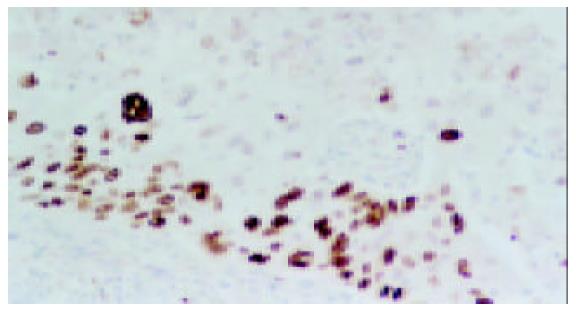
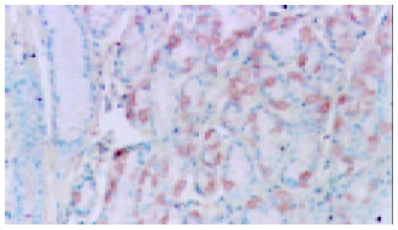
Figure 1 Immunohistochemical staining patterns of Ki-67 in gastric adenocarcinomas.
Positive nuclear staining in LSAB, × 200. Poorly-differentiated adenocarcinoma, demonstrated cell proliferative activity.
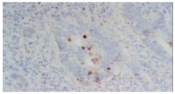
Figure 2 Apoptotic cancer cells in well-differentiated adeno-carcinoma detected by TUNEL method.
LSAB, × 200.
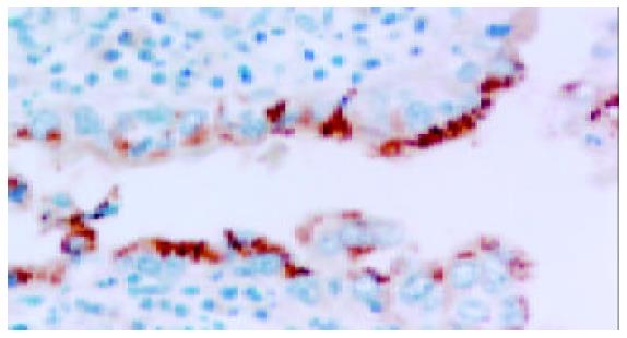
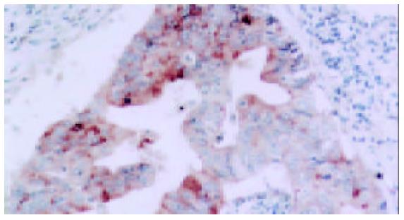
Figure 3 Strong Positive cytoplasma staining of EPR-1 in gastric adenocarcinomas (LSAB × 400).
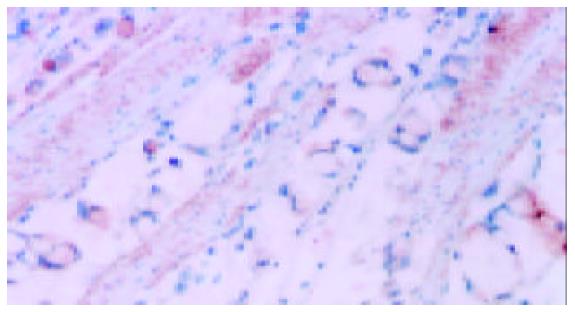
Figure 4 Positive staining of EPR-1 in signet-ring cell carcinoma invasion in smooth muscle and smooth muscle cells (LSAB × 200).
Figure 5 Moderate positive nuclear staining in benign gastric adenocytes (LSAB × 200).
Figure 6 Positive cytoplasma staining in well-differentiated lymph node metastasis (LSAB × 200).
Figure 7 Significant difference of AI and PI with EPR-1 score in primary adenocarcinoma.
A: The results of Pearson correlation coefficient, r = 0.296, P < 0.05; B: The results of pearson correlation coefficient, r = -0.204, P < 0.05 Weighted EPR-1 Score was positively correlated with apoptotic index, but negatively related with proliferative index in primary adenocarcinoma.
Figure 8 Significant difference of AI and PI with EPR-1 score in lymph node metastasis.
A: The results of pearson correlation coefficient, r = 0.332, P < 0.05; B: The results of pearson correlation coefficient, r = -0.336, P < 0.05. Weighted EPR-1 score is positively correlated with apoptosis index, but negatively related with proliferative index in lymph node metastasis.
- Citation: Yao XQ, Liu FK, Li JS, Qi XP, Wu B, Yin HL, Ma HH, Shi QL, Zhou XJ. Significance of effector protease receptor-1 expression and its relationship with proliferation and apoptotic index in patients with primary advanced gastric adenocarcinoma. World J Gastroenterol 2004; 10(9): 1262-1267
- URL: https://www.wjgnet.com/1007-9327/full/v10/i9/1262.htm
- DOI: https://dx.doi.org/10.3748/wjg.v10.i9.1262


