Copyright
©The Author(s) 2004.
World J Gastroenterol. Apr 1, 2004; 10(7): 985-990
Published online Apr 1, 2004. doi: 10.3748/wjg.v10.i7.985
Published online Apr 1, 2004. doi: 10.3748/wjg.v10.i7.985
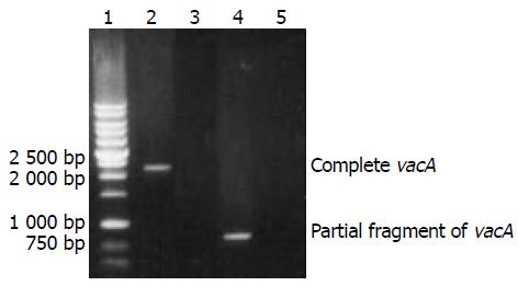
Figure 1 The target amplification products with whole length of vacA gene from H pylori strain NCTC11637 and partial frag-ment of vacA gene from a H pylori isolate.
Lane 1, DNA marker; lane 2, an amplification fragment of complete vacA gene from H pylori strain NCTC11637; lane 4, an amplification fragment of partial vacA gene from a H pylori isolate; and lanes 3 and 5, blank controls.
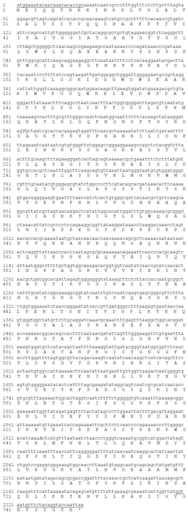
Figure 2 Nucleotide and putative amino acid sequences of vacA gene from H pylori strain NCTC 11637.
Note: Underlined areas indicate the position of primers; C, T, C and A are replaced by t, c, g and g in the n
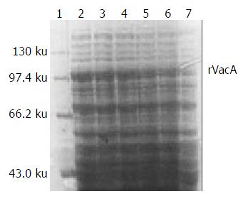
Figure 3 Expression of rVacA induced by IPTG at different concentrations.
Lane 1, the protein marker; lanes 2-4, IPTG at 1.0, 0.5, 0.1 mmol/L respectively; lanes 5 and 6, bacterial pre-cipitate and supernatant with IPTG at 0.5 mmol/L, respectively; and lane 7, the negative control.ucleotide sequence from strain NCTC 11637, respectively, but the encoded amino acid residuals are not altered (are the changes correct?). “*” means stop codon.
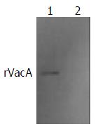
Figure 4 Western blot result of rabbit antibody against whole cell of H pylori and rVacA.
Lane 1, rVacA expressed by pET32a-vacA-E.coliBL21DE3; and lane 2: negative control of E.coliB L21DE3.
- Citation: Yan J, Mao YF. Construction of a prokaryotic expression system of vacA gene and detection of vacA gene, VacA protein in Helicobacter pylori isolates and ant-VacA antibody in patients’ sera. World J Gastroenterol 2004; 10(7): 985-990
- URL: https://www.wjgnet.com/1007-9327/full/v10/i7/985.htm
- DOI: https://dx.doi.org/10.3748/wjg.v10.i7.985


