Copyright
©The Author(s) 2004.
World J Gastroenterol. Mar 15, 2004; 10(6): 871-874
Published online Mar 15, 2004. doi: 10.3748/wjg.v10.i6.871
Published online Mar 15, 2004. doi: 10.3748/wjg.v10.i6.871
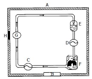
Figure 1 Preservation of continuously hypothermic machine perfusion.
A: Organ hypothermic preservation box, B: Tem-perature displayer, C: Perfusion pump, D: Perfusion pump, (Displaying the perfusion pressure). E: Perfusate container, F: Organ preservation container, G: pH displayer, H: Entry of wire, (Being sealed while preservation).
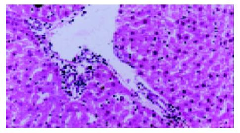
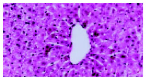
Figure 2 Histological and morphological findings in hypothermically preserved hepatocytes.
Group A: 24-h preservation, HE staining 100×.
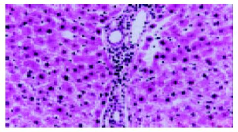
Figure 3 Histological and morphological findings in hypothermically preserved hepatocytes.
Group B: 24-h preservation, HE staining 100×.
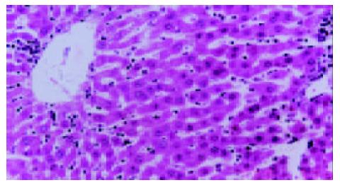
Figure 4 Histological and morphological findings in hypothermically preserved hepatocytes.
Group C: 24-h preservation, HE staining 100×.
Figure 5 Autoradiography of [-32P] ATP in hypothermically preserved hepatocytes.
Black spots in hepatocytes are the autoradiographies of [-32P] ATP, 4-h preservation, H E staining 100×.
- Citation: Tan XD, Egami H, Wang FS, Ogawa M. Protective effect of exogenous adenosine triphosphate on hypothermically preserved rat liver. World J Gastroenterol 2004; 10(6): 871-874
- URL: https://www.wjgnet.com/1007-9327/full/v10/i6/871.htm
- DOI: https://dx.doi.org/10.3748/wjg.v10.i6.871


