Copyright
©The Author(s) 2004.
World J Gastroenterol. Mar 15, 2004; 10(6): 795-799
Published online Mar 15, 2004. doi: 10.3748/wjg.v10.i6.795
Published online Mar 15, 2004. doi: 10.3748/wjg.v10.i6.795
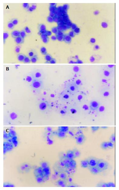
Figure 1 A: Untreated MGC803 Cells( apoptotic bodies could not be found), B: MGC803 cells treated with VCR 48 h (apoptotic bodies could be found), C: MGC803 cells treated with VCR+PD098059 48 h (apoptotic bodies could be found).
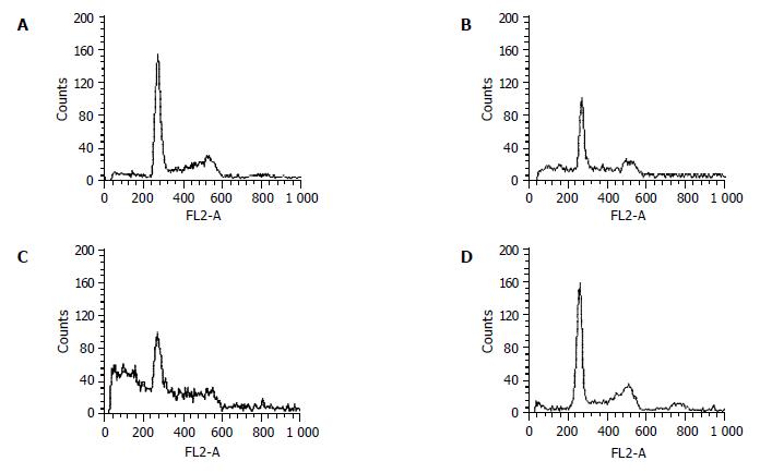
Figure 2 Cell cycle analysis of MGC803.
A: Untreated MGC803 cells at 72 h (G0G1: 48.20%; G2M: 28.18%, S: 14.16%, apoptosis: 8.46%), B: MGC803 cells treated with VCR (20 ng/mL) after 72 h (G0G1: 37.62%; G2M: 20.05%, S: 24.92%, apoptosis: 18.41%), C: MGC803 cells treated with VCR (20 ng/mL) + PD098059 (10 nmol/mL) after 72 h (G0G1: 32.53%; G2M: 13.91%, S: 18.95%, apoptosis: 35.61%), D: MGC803 cells treated with PD098059 (10 nmol/mL) only after 72 h (G0G1: 49.27%; G2M: 27.56%, S: 16.91%, apoptosis: 6.26%).
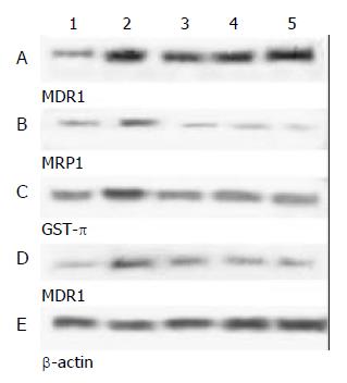
Figure 3 The expression of associated gene of MDR in MGC803 cells treated with VCR or VCR+PD098059 24h-72 h.
A: The expression of MDR1 of MGC803 cells treated with VCR increased. B,C: The expression of MRP1 and GST-π of MGC803 cells treated with VCR did not increase. D: The expression of MDR1 was inhibited when MGC803 cells treated with VCR and PD098059. E: The protein level of β-actin was detected to assess the loading amount in each well in SDS-PAGE. Lane 1, untreated cells; Lane 2, positive control cells; Lane 3, cells treated for 24 h; Lane 4, cells treated 48 h; Lane 5, cells treated for 72 h.
- Citation: Chen B, Jin F, Lu P, Lu XL, Wang PP, Liu YP, Yao F, Wang SB. Effect of mitogen-activated protein kinase signal transduction pathway on multidrug resistance induced by vincristine in gastric cancer cell line MGC803. World J Gastroenterol 2004; 10(6): 795-799
- URL: https://www.wjgnet.com/1007-9327/full/v10/i6/795.htm
- DOI: https://dx.doi.org/10.3748/wjg.v10.i6.795


