Copyright
©The Author(s) 2004.
World J Gastroenterol. Mar 1, 2004; 10(5): 699-702
Published online Mar 1, 2004. doi: 10.3748/wjg.v10.i5.699
Published online Mar 1, 2004. doi: 10.3748/wjg.v10.i5.699
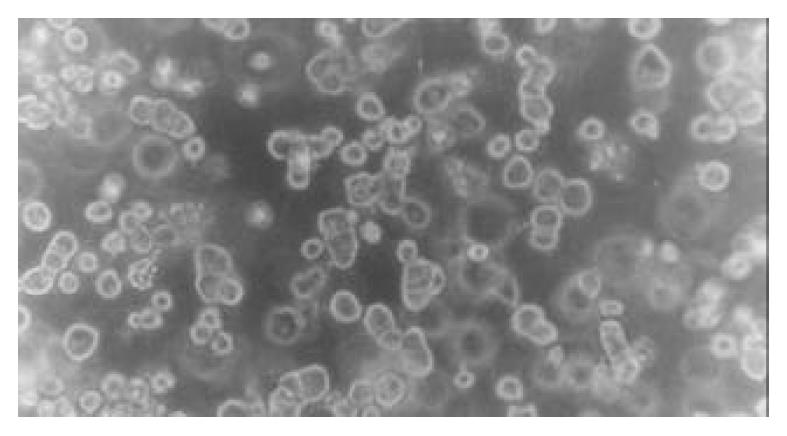
Figure 1 Morphology of rat hepatocytes in collagen gel mix-ture culture at 24 h (phase-contrast microscope ×200).
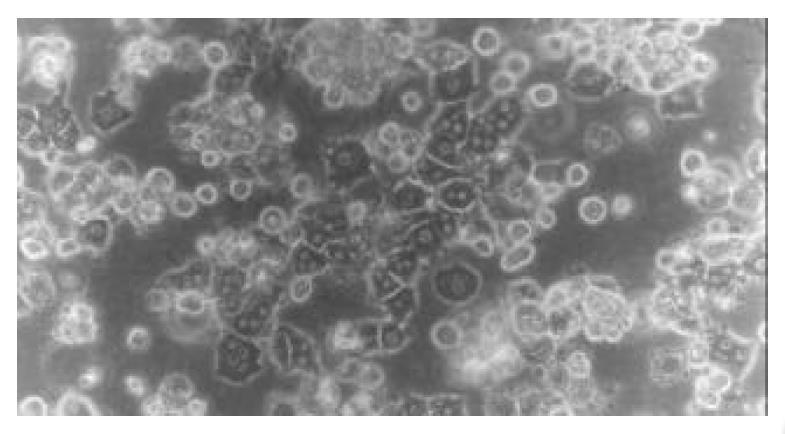
Figure 2 Morphology of rat hepatocytes in collagen gel mix-ture culture at 72 h (phase-contrast microscope ×200).
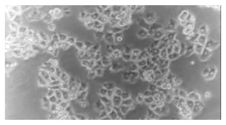
Figure 3 Morphology of piglet hepatocytes cultured on a single layer of collagen gel at 4 h (phase-contrast microscope ×200).
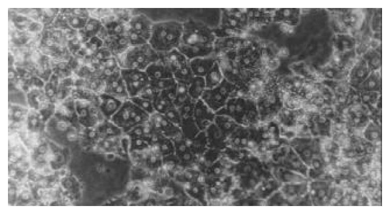
Figure 4 Morphology of piglet hepatocytes cultured in sand-wich configurations on d 5 (phase-contrast microscope ×200).
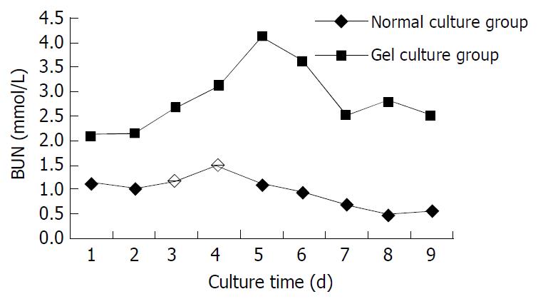
Figure 5 BUN level in supernatant of medium.
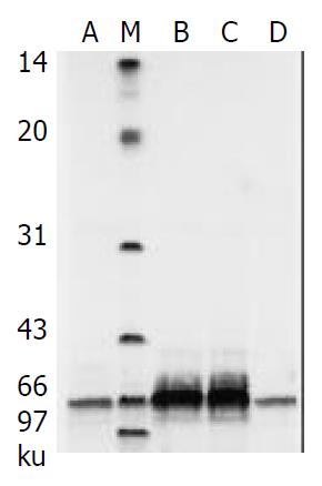
Figure 6 Albumin secretion in cultured piglet hepatocytes by SDS-PAGE analysis.
M: Low-range protein molecular weight markers; lanes A, B, C, D are culture media on days 1, 3, 5, 7.
- Citation: Wang YJ, Liu HL, Guo HT, Wen HW, Liu J. Primary hepatocyte culture in collagen gel mixture and collagen sandwich. World J Gastroenterol 2004; 10(5): 699-702
- URL: https://www.wjgnet.com/1007-9327/full/v10/i5/699.htm
- DOI: https://dx.doi.org/10.3748/wjg.v10.i5.699


