Copyright
©The Author(s) 2004.
World J Gastroenterol. Mar 1, 2004; 10(5): 626-629
Published online Mar 1, 2004. doi: 10.3748/wjg.v10.i5.626
Published online Mar 1, 2004. doi: 10.3748/wjg.v10.i5.626
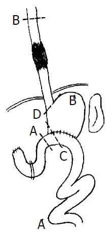
Figure 1 Resection range.
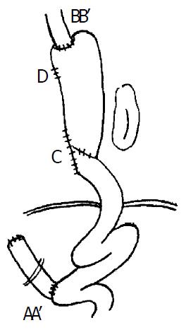
Figure 2 Postoperative situation.
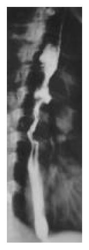
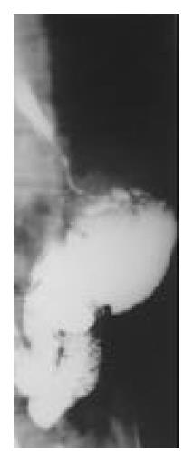
Figure 3 Preoperative barium.
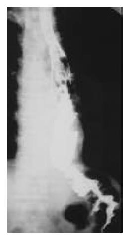
Figure 4 Preoperative barium meal examination, showing meal examination, showing re-mid thoracic esophageal tumor.
sidual stomach.
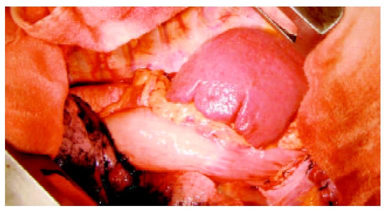
Figure 5 Field of operation, showing the spleen in the left tho-racic cavity.
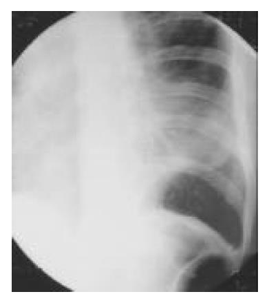
Figure 6 Postoperative roentgenogram of chest, showing the spleen shadow in the left thoracic cavity.
Figure 7 Postoperative barium meal examination, showing the esophagogastric anastomosis above the aortic arch, the residual stomach with previous gastrointestinal anastomosis, and the Roux-en-Y anastomosis.
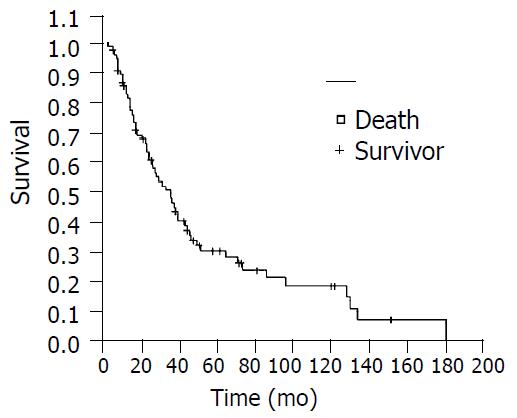
Figure 8 Kaplan-Meier survival curve.
- Citation: Chen YP, Yang JS, Liu DT, Chen YQ, Yang WP. Long-term effect on carcinoma of esophagus of distal subtotal gastrectomy. World J Gastroenterol 2004; 10(5): 626-629
- URL: https://www.wjgnet.com/1007-9327/full/v10/i5/626.htm
- DOI: https://dx.doi.org/10.3748/wjg.v10.i5.626


