Copyright
©The Author(s) 2004.
World J Gastroenterol. Feb 15, 2004; 10(4): 579-582
Published online Feb 15, 2004. doi: 10.3748/wjg.v10.i4.579
Published online Feb 15, 2004. doi: 10.3748/wjg.v10.i4.579
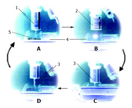
Figure 1 Procedures of paraffin-embed tissue micro-arraying.
(1. holing needle, 2. stereomicroscope lens, 3. sampling needle, 4. paraffin block-fixing box, 5. recipient paraffin block).
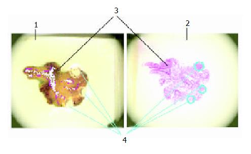
Figure 2 Defining accurate sampling sites in donor block un-der stereomicroscope.
(1. Paraffin donor block and paraffin embedded tissue under stereomicroscope, 2. Routine section and HE staining of paraffin embedded donor tissue under stereomicroscope, 3. Regions of stroma, less cells or areas with no tissue, 4. Sampling locus.).
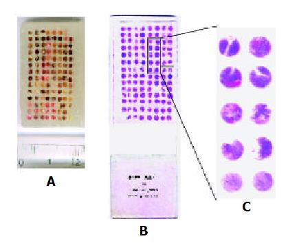
Figure 3 Pictures of paraffin-embedded tissue core arrange-ment (A), tissue spot arrays on slide (B) and magnified (X8) picture of selected array spots (C).
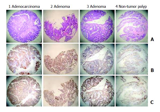
Figure 4 Photographs of different tissue array elements stained with HE(A) and immunohistochemistry (B: P53, C: PCNA).
- Citation: Dan HL, Zhang YL, Zhang Y, Wang YD, Lai ZS, Yang YJ, Cui HH, Jian YT, Geng J, Ding YQ, Guo CH, Zhou DY. A novel method for preparation of tissue microarray. World J Gastroenterol 2004; 10(4): 579-582
- URL: https://www.wjgnet.com/1007-9327/full/v10/i4/579.htm
- DOI: https://dx.doi.org/10.3748/wjg.v10.i4.579


