Copyright
©The Author(s) 2004.
World J Gastroenterol. Feb 15, 2004; 10(4): 540-544
Published online Feb 15, 2004. doi: 10.3748/wjg.v10.i4.540
Published online Feb 15, 2004. doi: 10.3748/wjg.v10.i4.540
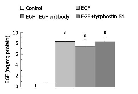
Figure 1 Levels of EGF in medium after incubation of SW480 cells with 0.
06% dimethyl sulfoxide (DMSO, the control group), 225 ng/mL (37.5 nmol/L) EGF in 0.6 mL/L DMSO, 225 ng/mL EGF + 2.5 μg/mL (17 nmol/L) EGF antibody in 0.6 mL/L DMSO, or 225 ng/mL EGF + 215 ng/mL (0.8 μmol/L) tyrphostin 51 in 0.6 mL/L DMSO serum-free medium for 48 h. Data are expressed as mean ± SD (n = 7). aP < 0.05 significantly different between the control and EGF-treated groups.
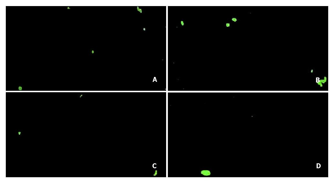
Figure 2 Apoptotic cells stained by annexin V-FITC binding (green) after incubation of SW480 cells with 0.
6 mL/L dimethyl sulfoxide (DMSO, the control group) (A), 225 ng/mL (37.5 nmol/L) EGF in 0.6 mL/L DMSO (B), 225 ng/mL EGF + 2.5 μg/mL (17 nmol/L) EGF antibody in 0.6 mL/L DMSO (C), or 225 ng/mL EGF + 215 ng/mL (0.8 μmol/L) tyrphostin 51 in 0.6 mL/L DMSO serum-free medium (D) for 12 h. MicrograpHmagnified by × 100 is the representative of seven independent experiments (n = 7).
Figure 3 Expression of phosphorylated EGF receptor (EGF-R) with the molecular weight of 145 kDa visualized by Western blotting (A) and quantitated by an image analysis system (B) after incubation of SW480 cells with 0.
6 mL/L dimethyl sulfoxide (DMSO, the control group), 225 ng/mL (37.5 nmol/L) EGF in 0.6 mL/L DMSO, 225 ng/mL EGF + 2.5 μg/mL (17 nmol/L) EGF antibody in 0.6 mL/L DMSO, or 225 ng/mL EGF + 215 ng/mL (0.8 μmol/L) tyrphostin 51 in 0.6 mL/L DMSO serum-free medium for 48 h. Samples were pooled from 7 independent experiments (n = 7). Density was calibrated by an internal control, α-tubulin (55 kDa).
Figure 4 Expression of p21 protein with the molecular weight of 21 kDa visualized by Western blotting (A) and quantitated by an image analysis system (B) after incubation of SW480 cells with 0.
6 mL/L dimethyl sulfoxide (DMSO, the control group), 225 ng/mL (37.5 nmol/L) EGF in 0.6 mL/L DMSO, 225 ng/mL EGF + 2.5 μg/mL (17 nmol/L) EGF antibody in 0.6 mL/L DMSO, or 225 ng/mL EGF + 215 ng/mL (0.8 μmol/L) tyrphostin 51 in 0.6 mL/L DMSO serum-free medium for 48 h. Samples were pooled from 7 independent experiments (n = 7). Density was calibrated by an internal control, α-tubulin (55 kDa).
- Citation: Chao JC, Peng WL, Chen SH. Effects of epidermal growth factor and its signal transduction inhibitors on apoptosis in human colorectal cancer cells. World J Gastroenterol 2004; 10(4): 540-544
- URL: https://www.wjgnet.com/1007-9327/full/v10/i4/540.htm
- DOI: https://dx.doi.org/10.3748/wjg.v10.i4.540


