Copyright
©The Author(s) 2004.
World J Gastroenterol. Feb 1, 2004; 10(3): 460-462
Published online Feb 1, 2004. doi: 10.3748/wjg.v10.i3.460
Published online Feb 1, 2004. doi: 10.3748/wjg.v10.i3.460
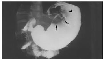
Figure 1 A large lobulated mass at the upper part of gastric body (arrow head) shown by radiography.
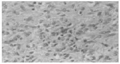
Figure 2 Packed spindle-shaped cells, proliferated vessels and inflammatory cells, mostly eosinophils observed in tumor (H&E, 10 × 40).
- Citation: Chongsrisawat V, Yimyeam P, Wisedopas N, Viravaidya D, Poovorawan Y. Unusual manifestations of gastric inflammatory fibroid polyp in a child. World J Gastroenterol 2004; 10(3): 460-462
- URL: https://www.wjgnet.com/1007-9327/full/v10/i3/460.htm
- DOI: https://dx.doi.org/10.3748/wjg.v10.i3.460


