Copyright
©The Author(s) 2004.
World J Gastroenterol. Feb 1, 2004; 10(3): 337-341
Published online Feb 1, 2004. doi: 10.3748/wjg.v10.i3.337
Published online Feb 1, 2004. doi: 10.3748/wjg.v10.i3.337
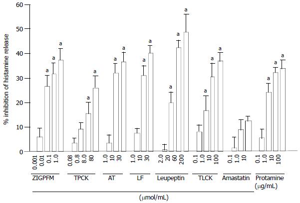
Figure 1 Inhibition of anti-IgE (10 µg/mL) induced histamine release from dispersed colon mast cells by the protease inhibitors.
The inhibitors and anti-IgE were added to cells at the same time (no preincubation). Data are presented as mean ± SEM for four to six separate experiments performed in duplicate. aP < 0.05 compared with the responses with uninhibited controls. AT = α1-antitrypsin; LF = lactoferrin.
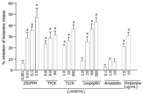
Figure 2 Inhibition of anti-IgE (10 µg/mL) induced histamine release from dispersed colon mast cells by the protease inhibitors.
The inhibitors were preincubated with cells for 20 min before anti-IgE was added. Data are presented as mean ± SEM for four to six separate experiments performed in duplicate. aP < 0.05 compared with the responses with uninhibited controls.
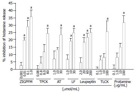
Figure 3 Inhibition of calcium ionophore (1 µg/mL) induced histamine release from dispersed colon mast cells by the protease inhibitors.
The inhibitors and anti-IgE were added to cells at the same time (no preincubation). Data are presented as mean ± SEM for four to six separate experiments performed in duplicate. aP < 0.05 compared with the responses with uninhibited controls. AT = α1-antitrypsin; LF = lactoferrin.
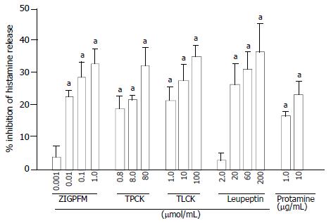
Figure 4 Inhibition of calcium ionophore (1 µg/mL) induced histamine release from dispersed colon mast cells by the protease inhibitors.
The inhibitors were preincubated with cells for 20 min before calcium ionophore being added. Data are presented as mean ± SEM for four to six separate experiments performed in duplicate. aP < 0.05 compared with the responses with uninhibited controls.
- Citation: He SH, Xie H. Modulation of histamine release from human colon mast cells by protease inhibitors. World J Gastroenterol 2004; 10(3): 337-341
- URL: https://www.wjgnet.com/1007-9327/full/v10/i3/337.htm
- DOI: https://dx.doi.org/10.3748/wjg.v10.i3.337


