Copyright
©The Author(s) 2004.
World J Gastroenterol. Jan 15, 2004; 10(2): 200-204
Published online Jan 15, 2004. doi: 10.3748/wjg.v10.i2.200
Published online Jan 15, 2004. doi: 10.3748/wjg.v10.i2.200
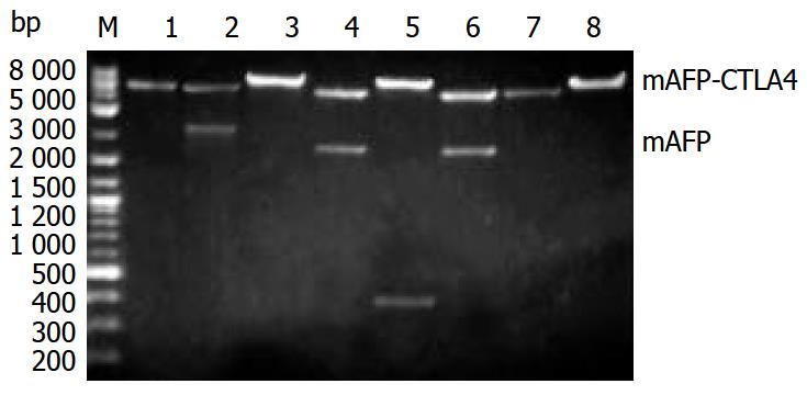
Figure 1 Identification of pmAFP and pmAFP-CTLA4 with restriction enzyme analysis.
M: DNA marker, Lane 1: pcDNA3.1/ EcoRI, Lane 2: pmAFP-CTLA4/EcoRI + XbaI, Lane 3: pmAFP-CTLA4/EcoRI, Lane 4: pmAFP-CTLA4/EcoRI+XhoI, Lane 5: pmAFP-CTLA4/XhoI + XbaI, Lane 6: pmAFP/EcoRI + XbaI, Lane 7: pcDNA3.1/EcoRI, Lane 8: pmAFP/EcoRI.
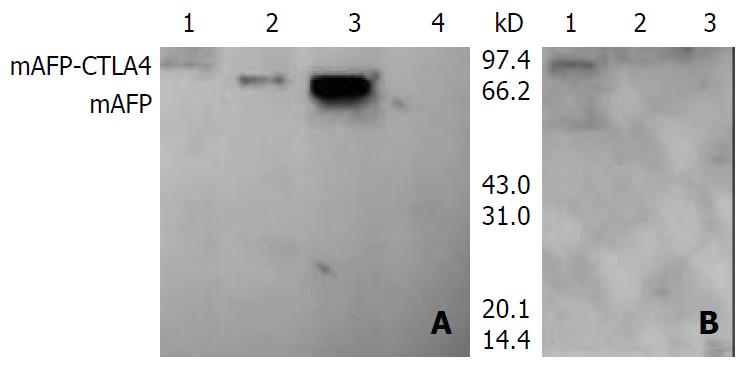
Figure 2 Detection of protein expression of plasmids by West-ern blotting.
A: the blot was probed with anti-AFP. Lane 1: CHO/pmAFP-CTLA, Lane 2: CHO/pmAFP, Lane 3: Hepa 1-6, Lane 4: CHO/pcDNA3.1, B: the blot was probed with anti-CTLA4. Lane 1: CHO/pmAFP-CTLA, Lane 2: CHO/pmAFP, Lane 3: CHO/pcDNA3.1.
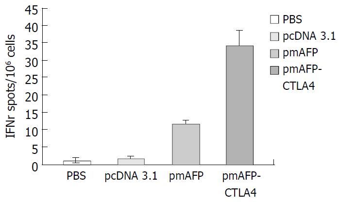
Figure 3 Number of IFN-γ-producing cells in 4 different groups
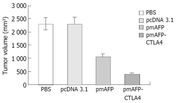
Figure 4 Comparison of tumor masses in 4 groups on 22 days following tumor challenge.
- Citation: Tian G, Yi JL, Xiong P. Antitumor immunopreventive effect in mice induced by DNA vaccine encoding a fusion protein of α-fetoprotein and CTLA4. World J Gastroenterol 2004; 10(2): 200-204
- URL: https://www.wjgnet.com/1007-9327/full/v10/i2/200.htm
- DOI: https://dx.doi.org/10.3748/wjg.v10.i2.200


