Copyright
©The Author(s) 2004.
World J Gastroenterol. Sep 15, 2004; 10(18): 2652-2656
Published online Sep 15, 2004. doi: 10.3748/wjg.v10.i18.2652
Published online Sep 15, 2004. doi: 10.3748/wjg.v10.i18.2652
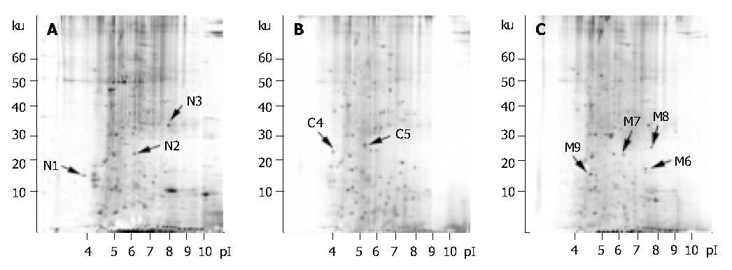
Figure 1 Silver-stained two-dimensional electrophoretic images of hydrophobic proteins from (A) Normal colon mucosa, (B) Primary colon cancer lesion, (C) Hepatic metastasis.
Figure 2 MALDI-TOF mass spectrometry and peptide mass fingerprint analysis of the differential protein spots (A) N2 protein spot from normal colon mucosa, (B) M6 protein spot from hepatic metastasis.
- Citation: Yu B, Li SY, An P, Zhang YN, Liang ZJ, Yuan SJ, Cai HY. Comparative study of proteome between primary cancer and hepatic metastatic tumor in colorectal cancer. World J Gastroenterol 2004; 10(18): 2652-2656
- URL: https://www.wjgnet.com/1007-9327/full/v10/i18/2652.htm
- DOI: https://dx.doi.org/10.3748/wjg.v10.i18.2652


