Copyright
©The Author(s) 2004.
World J Gastroenterol. Aug 15, 2004; 10(16): 2444-2446
Published online Aug 15, 2004. doi: 10.3748/wjg.v10.i16.2444
Published online Aug 15, 2004. doi: 10.3748/wjg.v10.i16.2444
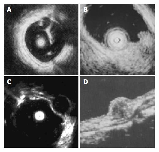
Figure 1 EUS imagines of normal wall and SMT of the large intestine.
A: The normal wall was displayed in 5 layers; B: Li-poma imagine showed a hyperechoic homogeneous mass lo-cated in the third layer; C: Leiomyoma imagine showed a hypoechoic homogeneous mass originated from the 4th layer; D: Rectal carcinoid imagine showed a submucosal hypoechoic mass with a homogenous echo.
- Citation: Zhou PH, Yao LQ, Zhong YS, He GJ, Xu MD, Qin XY. Role of endoscopic miniprobe ultrasonography in diagnosis of submucosal tumor of large intestine. World J Gastroenterol 2004; 10(16): 2444-2446
- URL: https://www.wjgnet.com/1007-9327/full/v10/i16/2444.htm
- DOI: https://dx.doi.org/10.3748/wjg.v10.i16.2444


