Copyright
©The Author(s) 2004.
World J Gastroenterol. Jul 15, 2004; 10(14): 2147-2149
Published online Jul 15, 2004. doi: 10.3748/wjg.v10.i14.2147
Published online Jul 15, 2004. doi: 10.3748/wjg.v10.i14.2147
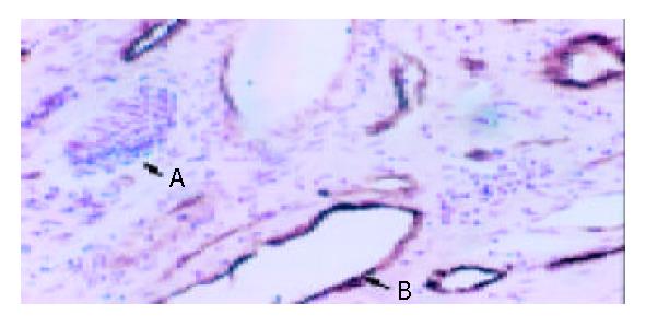
Figure 1 Immunohistochemical staining with anti-F VIII-RAg (S-P, original magnification: × 400).
A indicates the lymphatic vessel endothelium. B indicates the brown venous endothelium.
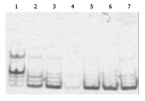
Figure 2 Telomerase activity of HCC tissues.
Lane 1: Positive control. Lanes 2, 3: HCC tissue with low differentiation with a 6-bp interval ladder pattern. Lane 4: No ladder in a djacent normal tissues. Lanes 5, 6: HCC tissue with high differentiation with a 6-bp interval ladder pattern. Lane 7: HCC with moderate differentiation with a 6-bp interval ladder pattern.
- Citation: Piao YF, He M, Shi Y, Tang TY. Relationship between microvessel density and telomerase activity in hepatocellular carcinoma. World J Gastroenterol 2004; 10(14): 2147-2149
- URL: https://www.wjgnet.com/1007-9327/full/v10/i14/2147.htm
- DOI: https://dx.doi.org/10.3748/wjg.v10.i14.2147


