Copyright
©The Author(s) 2004.
World J Gastroenterol. Jul 1, 2004; 10(13): 1984-1988
Published online Jul 1, 2004. doi: 10.3748/wjg.v10.i13.1984
Published online Jul 1, 2004. doi: 10.3748/wjg.v10.i13.1984
Figure 1 HE staining of gastric carcinoma.
A: well differentiated gastric carcinoma; B: Moderately differentiated gastric carcinoma; C: Poorly differentiated gastric carcinoma (Original magnification: × 200).
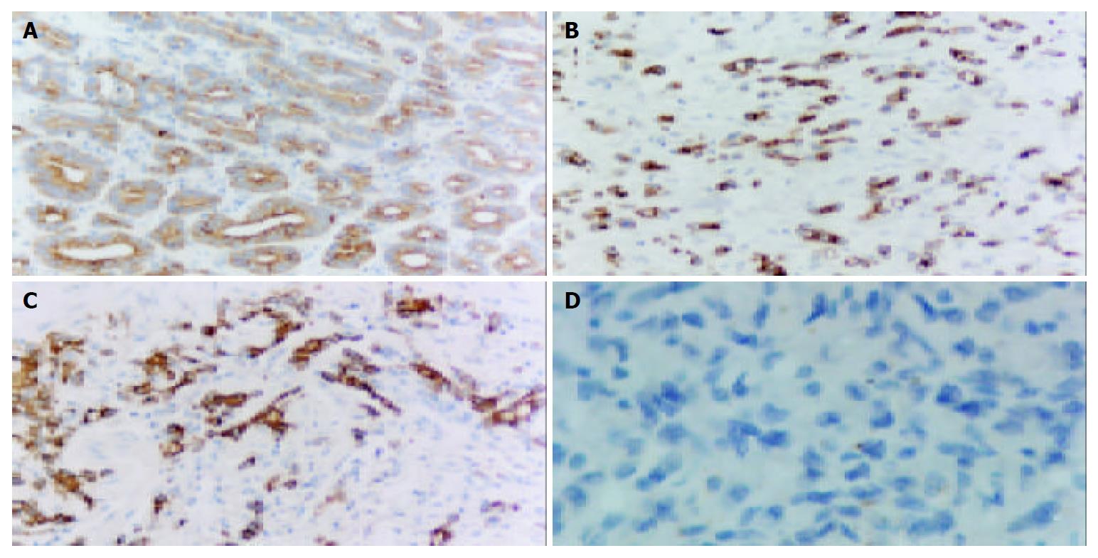
Figure 2 Survivin expression in gastric carcinoma.
A: well differentiated gastric carcinoma; B: Moderately differentiated gastric carcinoma; C: Poorly differentiated gastric carcinoma; D: Substitution for antibody with PBS as negative control (Original magnification: × 200).
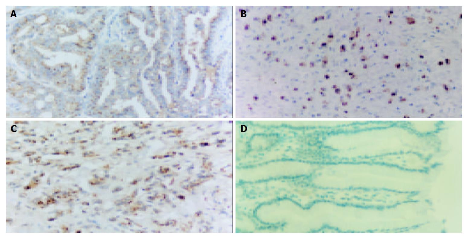
Figure 3 Expression of caspase-3 in gastric carcinoma.
A: well differentiated gastric carcinoma; B: Moderately differentiated gastric carcinoma; C: Poorly differentiated gastric carcinoma; D: Substitution for antibody with PBS as negative control (Original magnification: × 200).
Figure 4 Apoptosis in gastric carcinoma.
A: Positive; B: Substitution for terminal deoxynucleotidyl transferase with distilled water as negative control (Original magnification: × 200).
- Citation: Li YH, Wang C, Meng K, Chen LB, Zhou XJ. Influence of survivin and caspase-3 on cell apoptosis and prognosis in gastric carcinoma. World J Gastroenterol 2004; 10(13): 1984-1988
- URL: https://www.wjgnet.com/1007-9327/full/v10/i13/1984.htm
- DOI: https://dx.doi.org/10.3748/wjg.v10.i13.1984


