Copyright
©The Author(s) 2004.
World J Gastroenterol. Jul 1, 2004; 10(13): 1867-1871
Published online Jul 1, 2004. doi: 10.3748/wjg.v10.i13.1867
Published online Jul 1, 2004. doi: 10.3748/wjg.v10.i13.1867
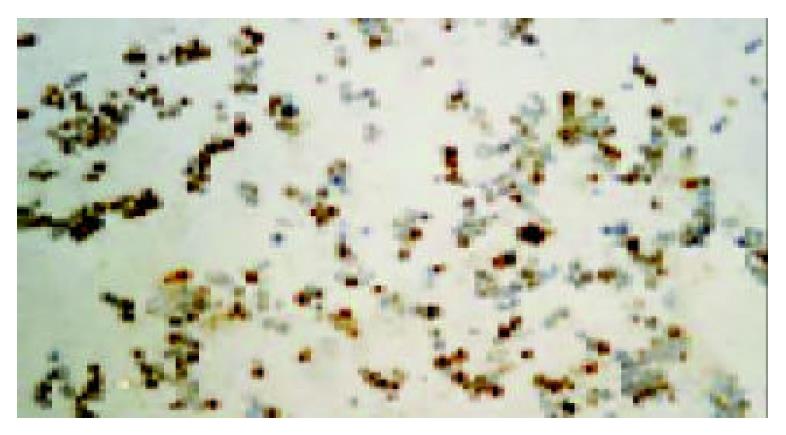
Figure 1 Immunohistochemistry analysis for endostatin pro-tein in vitro (× 100).
Extensive positive staining was observed in the cytoplasm of transduced cells. This indicated that human endostatin gene mediated by recombinant adenovirus was highly expressed in BEL-7402 cells.
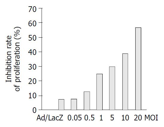
Figure 2 Effect of Ad/hEndo on the proliferation of HUVEC cells.
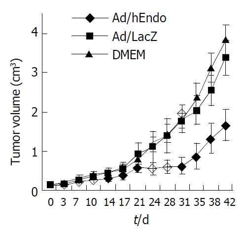
Figure 3 Effect of Ad/hEndo on growth of BEL-7402 xenografted liver tumors.
Figure 4 Northern blotting of endostatin mRNA in vivo.
Lanes 1-3: RNA extracted from tumors on d 1, 4 and 8 after Ad/hEndo injection, respectively; Lane 4: RNA extracted from control tumors injected with DMEM.
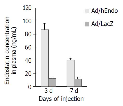
Figure 5 Detection of endostatin concentration in plasma by ELISA.
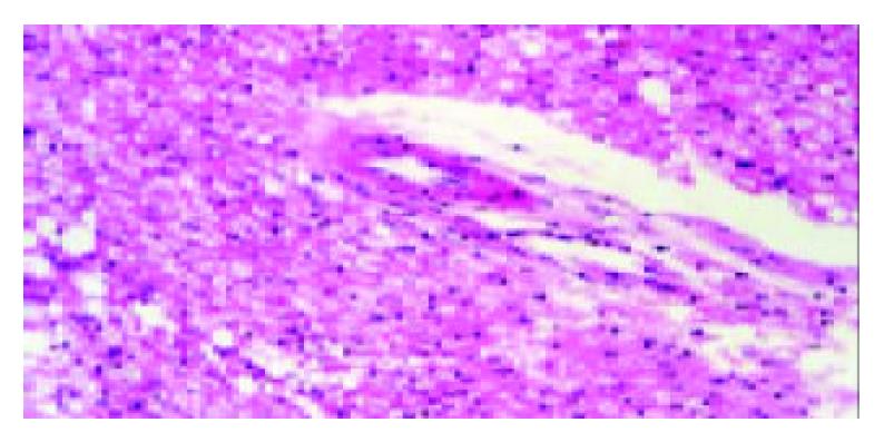
Figure 6 Pathological changes of liver 1 wk after 6 wk of treatment with Ad/hEndo (HE, original magnification: × 200).
- Citation: Li L, Huang JL, Liu QC, Wu PH, Liu RY, Zeng YX, Huang WL. Endostatin gene therapy for liver cancer by a recombinant adenovirus delivery. World J Gastroenterol 2004; 10(13): 1867-1871
- URL: https://www.wjgnet.com/1007-9327/full/v10/i13/1867.htm
- DOI: https://dx.doi.org/10.3748/wjg.v10.i13.1867


