Copyright
©The Author(s) 2004.
World J Gastroenterol. Jun 15, 2004; 10(12): 1841-1843
Published online Jun 15, 2004. doi: 10.3748/wjg.v10.i12.1841
Published online Jun 15, 2004. doi: 10.3748/wjg.v10.i12.1841
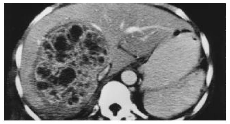
Figure 1 Contrast CT scan showing heterogeneous contrast enhancement in the lesion with a multi-septated appearance.
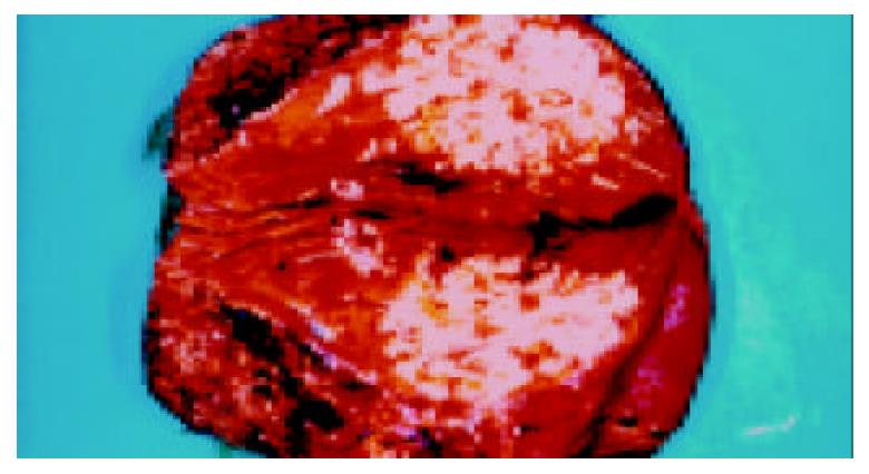
Figure 2 Yellowish firming well-circumscribed mass in the right hepatectomy specimen.
- Citation: Lo OS, Poon RT, Lam CM, Fan ST. Inflammatory pseudotumor of the liver in association with a gastrointestinal stromal tumor: A case report. World J Gastroenterol 2004; 10(12): 1841-1843
- URL: https://www.wjgnet.com/1007-9327/full/v10/i12/1841.htm
- DOI: https://dx.doi.org/10.3748/wjg.v10.i12.1841


