Copyright
©The Author(s) 2004.
World J Gastroenterol. Jun 1, 2004; 10(11): 1583-1588
Published online Jun 1, 2004. doi: 10.3748/wjg.v10.i11.1583
Published online Jun 1, 2004. doi: 10.3748/wjg.v10.i11.1583
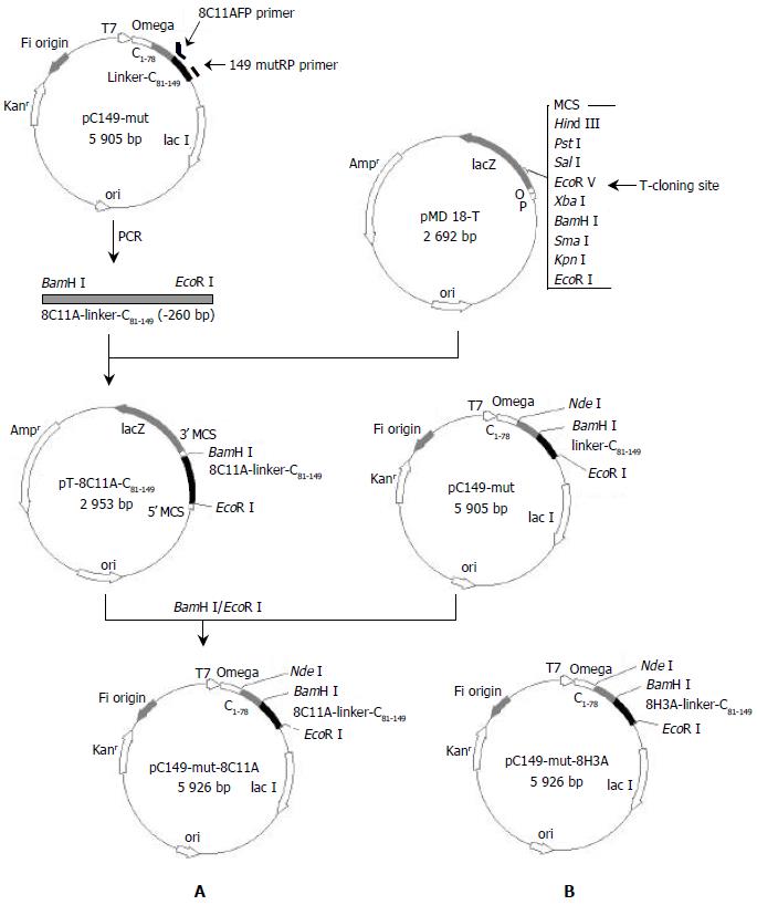
Figure 1 Expression vector construction of pC149-mut-8C11A and pC149-mut-8H3A.
A: Construction of pC149-mut-8C11A, B: Map of pC149-mut-8H3A, which was constructed as pC149-mut-8C11A except using the primer 8H3AFP instead of 8C11AFP.
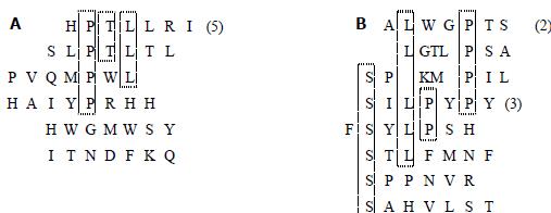
Figure 2 Sequences of peptides selected by monoclonal antibodies.
A: Selected by mAb 8C11, B: Selected by mAb 8H3.
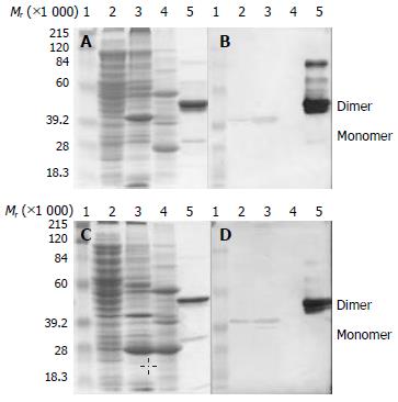
Figure 3 Expression and Western blotting of pC149-mut-8C11A and pC149-mut-8H3A.
A, C: SDS-PAGE, B, D: Western blot-ting of mAbs 8C11 and 8H3 respectively, A, B: pC149-mut-8C11A, C, D: pC149-mut-8H3A. 1, Protein molecular weight marker; 2, Supernatant of ultrasonic lysate; 3, Deposition of ultrasonic lysate; 4, C149-mut control; 5, Purified NE2 antigen
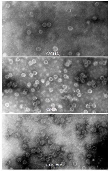
Figure 4 Virus like particles assemblied by recombinant protein C8C11A, C8H3A or C149 mut (Negative staining electron microscopy, × 100000).
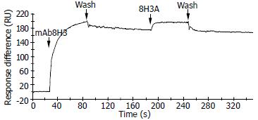
Figure 5 Binding curve of chemo-synthesized seven peptide 8H3A against monoclonal antibody 8H3 in BIAcore biosensor.
- Citation: Gu Y, Zhang J, Wang YB, Li SW, Yang HJ, Luo WX, Xia NS. Selection of a peptide mimicking neutralization epitope of hepatitis E virus with phage peptide display technology. World J Gastroenterol 2004; 10(11): 1583-1588
- URL: https://www.wjgnet.com/1007-9327/full/v10/i11/1583.htm
- DOI: https://dx.doi.org/10.3748/wjg.v10.i11.1583


