Copyright
©The Author(s) 1995.
World J Gastroenterol. Oct 1, 1995; 1(1): 60-61
Published online Oct 1, 1995. doi: 10.3748/wjg.v1.i1.60
Published online Oct 1, 1995. doi: 10.3748/wjg.v1.i1.60
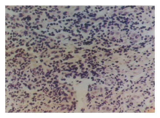
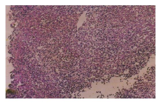
Figure 1 Inflammation granular tissue (HE × 200).
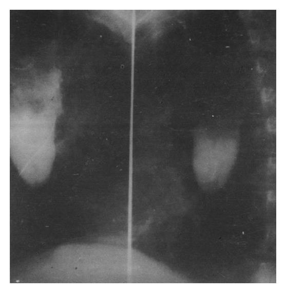
Figure 2 Esophageal obstruction piace is blind and no mucosa destroyed.
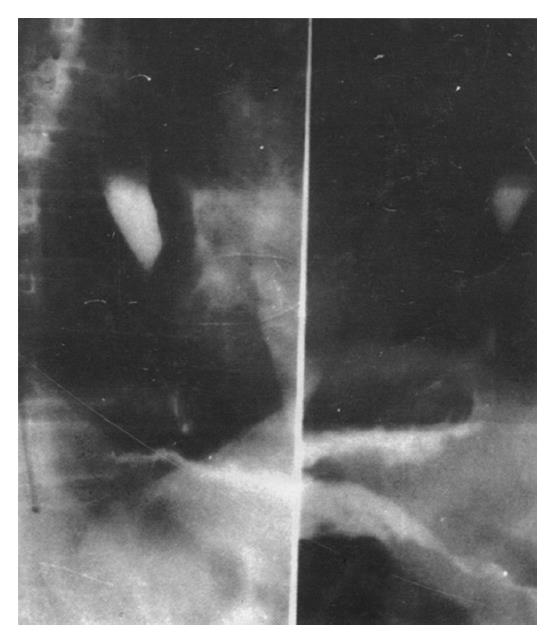
Figure 3 Esophageal intubation injection of contrast medium.
There is a line-like stenosis blow the obstruction.
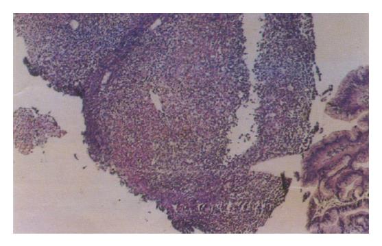
Figure 4 Local part seems to be a crack-like ulcer (HE × 45).
Figure 5 Local part seems to be a crack-like ulcer (HE × 75).
- Citation: Yu GH, Su ZG, Zou J, Wang YB. Esophageal Crohn's disease: A case report. World J Gastroenterol 1995; 1(1): 60-61
- URL: https://www.wjgnet.com/1007-9327/full/v1/i1/60.htm
- DOI: https://dx.doi.org/10.3748/wjg.v1.i1.60


