Copyright
©The Author(s) 1995.
World J Gastroenterol. Oct 1, 1995; 1(1): 43-47
Published online Oct 1, 1995. doi: 10.3748/wjg.v1.i1.43
Published online Oct 1, 1995. doi: 10.3748/wjg.v1.i1.43
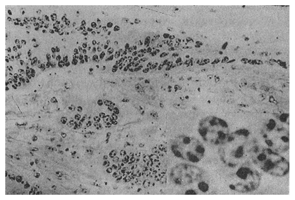
Figure 1 Moderately differentiated tubular adenomas (survived case) 10 × 20, 10 × 100).
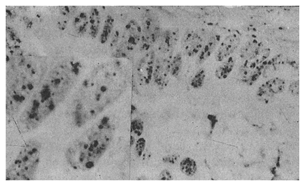
Figure 2 Moderately differentiated tubular adenomas (died case).
The AgNOR numbers, abnormal dots, large and small dots, and scattered and mixed types are significantly increased compared with survived cases (10 × 40, 10 × 100). AgNOR: Silver-stained nucleolar organizing regions.
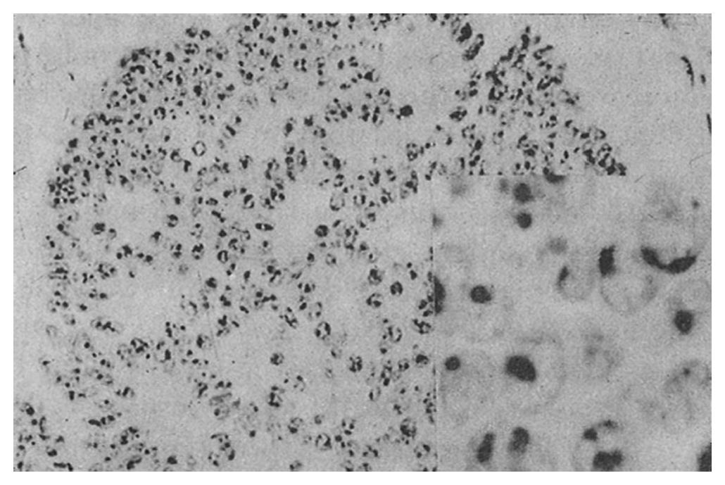
Figure 3 Moderately differentiated tubular adenomas (primary cancer of colon): 10 × 20, 10 × 100).
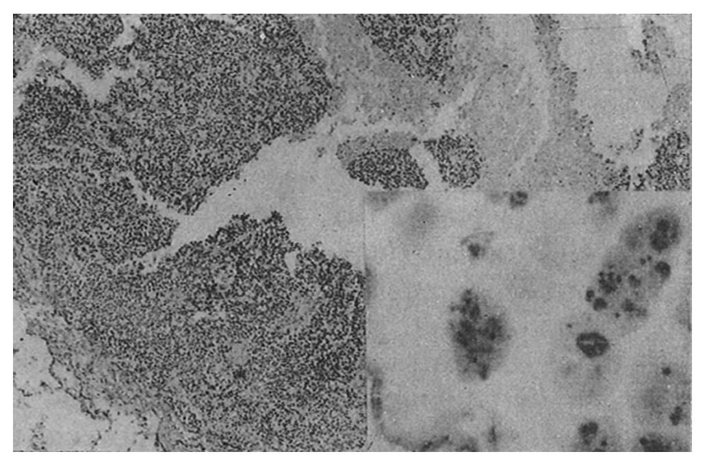
Figure 4 Moderately differentiated tubular adenomas (lymphonodic metastasis).
Compared with the primary cancer, the AgNOR number, large and small dots, and scattered type are increased significantly (10 × 10, 10 × 100); AgNOR: Silver-stained nucleolar organizing regions.
- Citation: Zhou ZF, Yuan SZ. Prognostic value of silver-stained nucleolar organizer regions in colorectal carcinoma. World J Gastroenterol 1995; 1(1): 43-47
- URL: https://www.wjgnet.com/1007-9327/full/v1/i1/43.htm
- DOI: https://dx.doi.org/10.3748/wjg.v1.i1.43


