
















©2014 Baishideng Publishing Group Inc.
World J Clin Cases. Jul 16, 2014; 2(7): 279-283
Published online Jul 16, 2014. doi: 10.12998/wjcc.v2.i7.279
Published online Jul 16, 2014. doi: 10.12998/wjcc.v2.i7.279
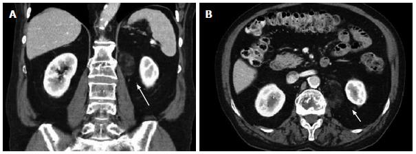
Figure 1 Initial computed tomography scan.
Shows incidentally found non-enhancing heterogeneous mass measuring approximately 3.8 cm × 2.3 cm in longitudinal (A) and anterior-posterior dimensions (B), just inferior to the left renal vein (long arrow) and medial to the left kidney (arrow).
- Citation: Hajiran A, Morley C, Jansen R, Kandzari S, Bacaj P, Zaslau S, Cardinal J. Perirenal extra-adrenal myelolipoma. World J Clin Cases 2014; 2(7): 279-283
- URL: https://www.wjgnet.com/2307-8960/full/v2/i7/279.htm
- DOI: https://dx.doi.org/10.12998/wjcc.v2.i7.279