
















©The Author(s) 2023.
World J Clin Cases. May 16, 2023; 11(14): 3340-3350
Published online May 16, 2023. doi: 10.12998/wjcc.v11.i14.3340
Published online May 16, 2023. doi: 10.12998/wjcc.v11.i14.3340
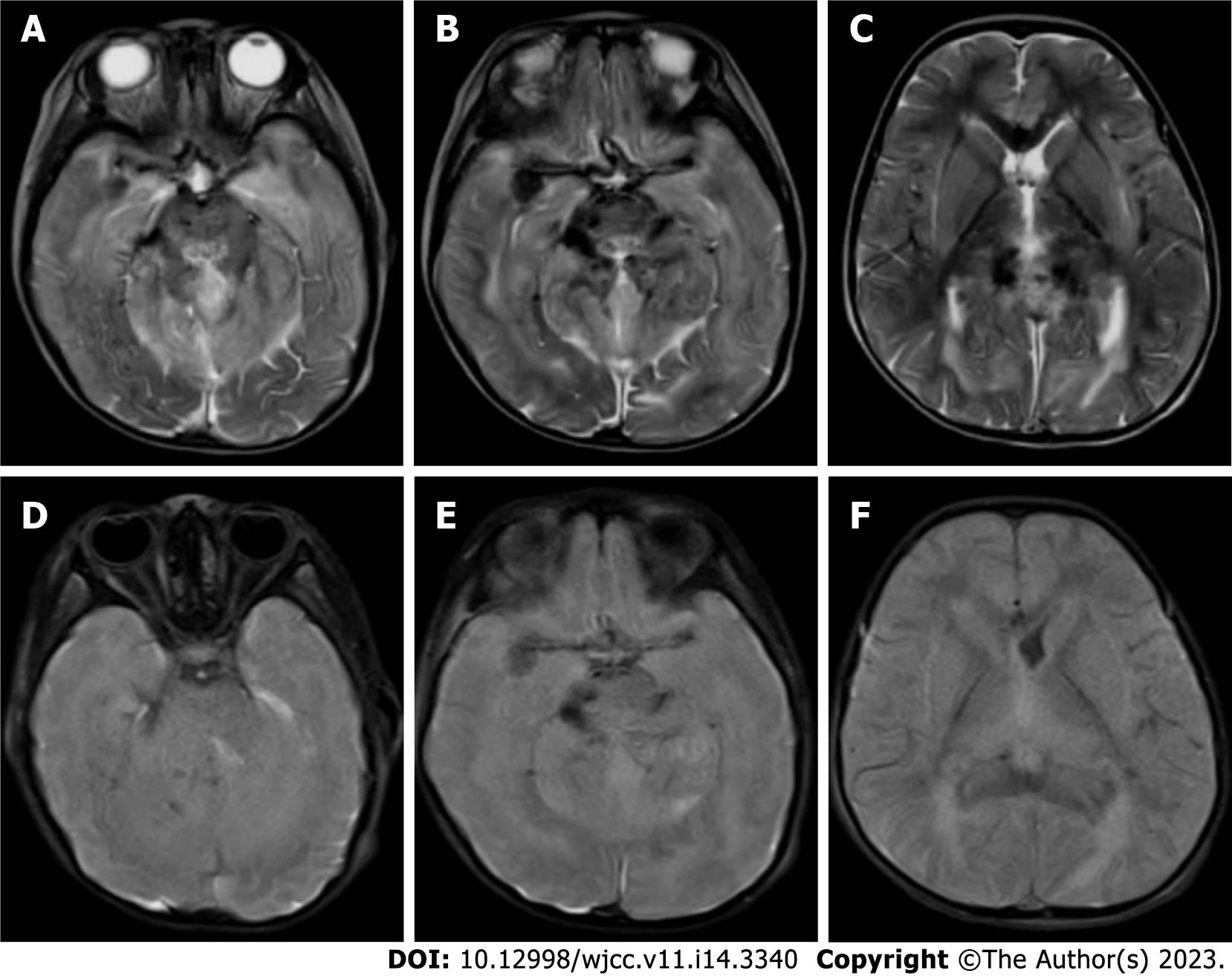
Figure 7 The imaging findings of head magnetic resonance on day 26 after admission.
A-C: T2-weighted imaging showed blurred boundary between gray matter and white matter, structural disorder of brain stem and cerebellar hemisphere, and multiple long T2 signal shadows; D-F: Fluid-attenuated inversion-recovery (FLAIR) images showed blurred gray and white matter, disordered structure, high FLAIR signal around the cerebellum and lateral ventricle, and significant narrowing of the lateral ventricle.
- Citation: Ding L, Huang TT, Ying GH, Wang SY, Xu HF, Qian H, Rahman F, Lu XP, Guo H, Zheng G, Zhang G. De novo mutation of NAXE (APOAIBP)-related early-onset progressive encephalopathy with brain edema and/or leukoencephalopathy-1: A case report. World J Clin Cases 2023; 11(14): 3340-3350
- URL: https://www.wjgnet.com/2307-8960/full/v11/i14/3340.htm
- DOI: https://dx.doi.org/10.12998/wjcc.v11.i14.3340