Copyright
©The Author(s) 2022.
World J Clin Cases. Jun 16, 2022; 10(17): 5877-5883
Published online Jun 16, 2022. doi: 10.12998/wjcc.v10.i17.5877
Published online Jun 16, 2022. doi: 10.12998/wjcc.v10.i17.5877
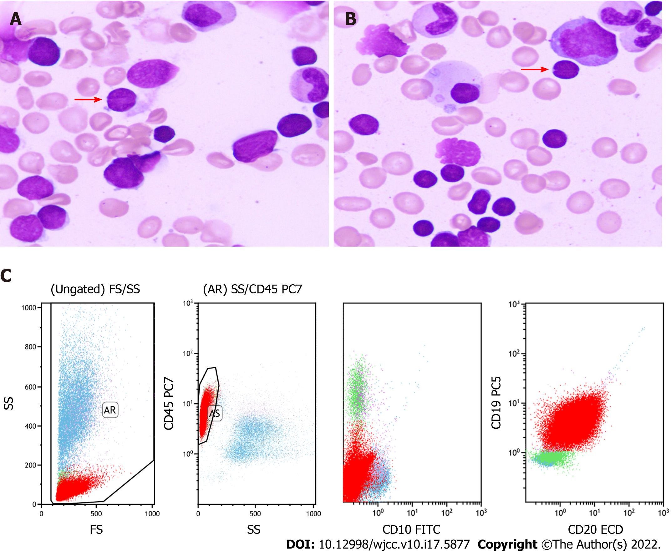
Figure 1 Morphology and flow cytometry analysis of the bone marrow.
A and B: The bone marrow (BM) was mainly infiltrated by monocytoid cells, accompanied with a few cells with cleaved or notched nuclei (pointed by arrows) [400 × (A) and 400 × (B)]; C: Flow cytometry revealed that the neoplastic cells (the red group) in BM were positive for CD19 and CD20 but negative for CD5 and CD10.
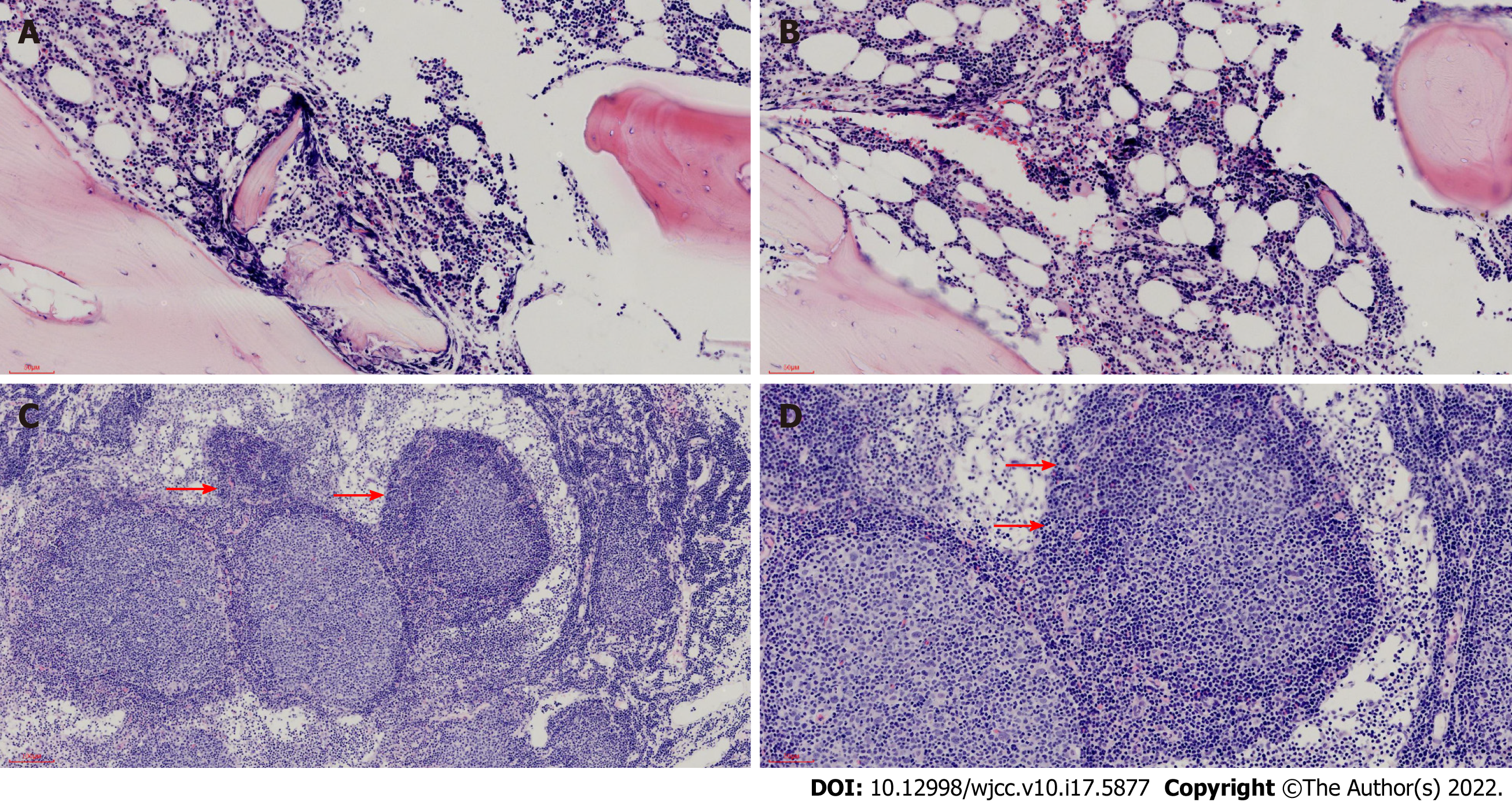
Figure 2 Morphology of the bone marrow and lymph node.
A and B: Hematoxylin and eosin (HE) stain displayed that the bone marrow was infiltrated by neoplastic lymphocytes in a paratrabecular pattern [100 × (A) and 200 × (B)]; C and D: HE stain showed that expanded follicles were infiltrated by neoplastic centroblasts and centrocytes, and serried monocytoid cells (arrows) [100 × (C) and 200 × (D)] surrounded them.
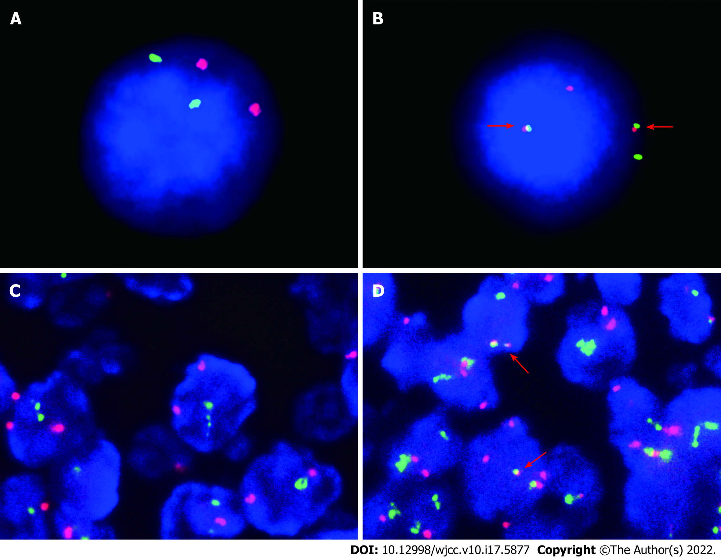
Figure 3 Fluorescence in situ hybridization analysis of the bone marrow and lymph node.
A-D: A and C were the control groups [1000 × (A) and 1000 × (B), 400 × (C) and 400 × (D)], the rearrangement of BCL-2/IGH (fusion of the red light and the green light) (B) was detected in the bone marrow and in the lymph node (fusion of the red light and the green light) (D) shown by fluorescence in situ hybridization.
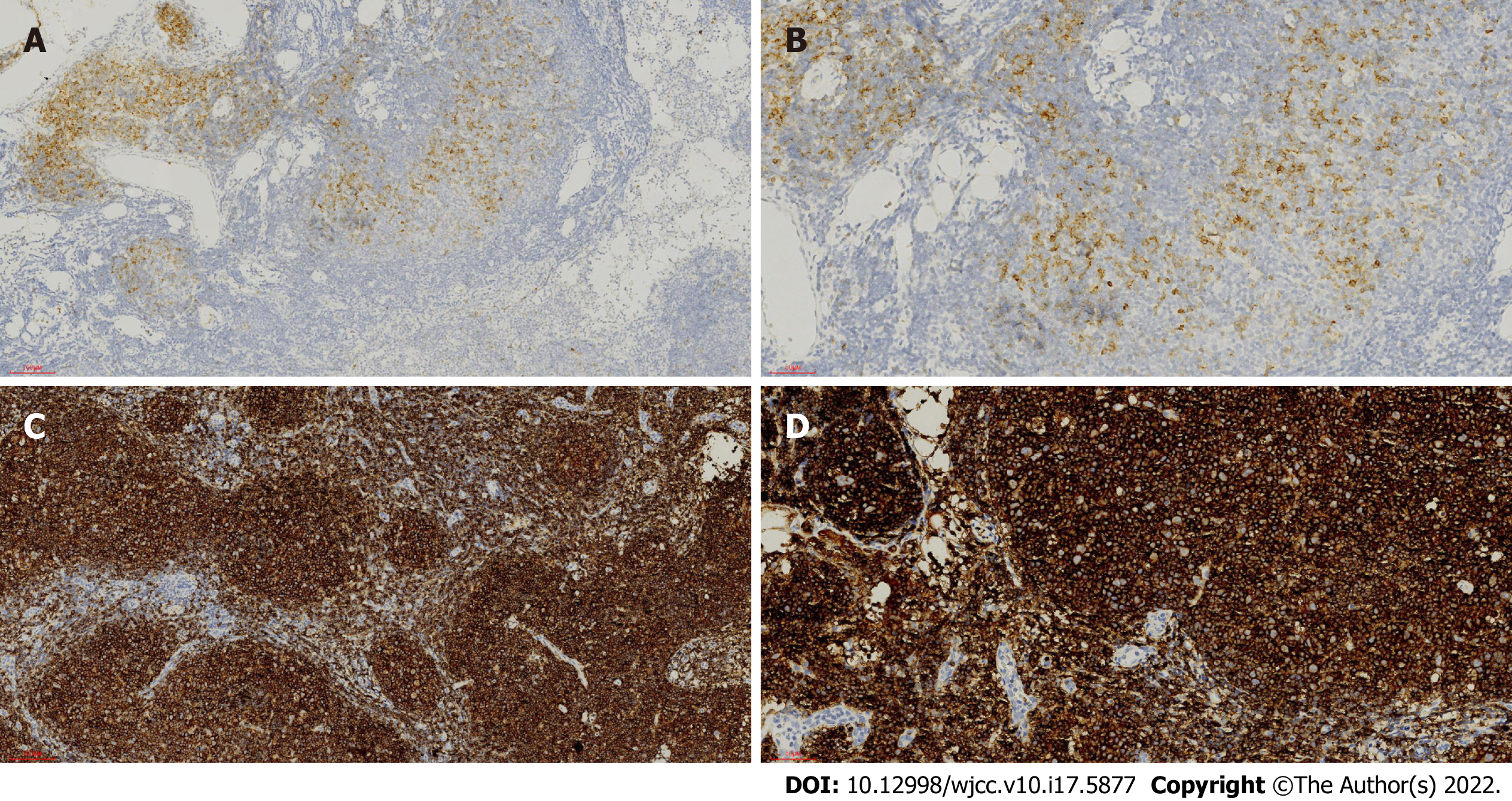
Figure 4 Immunohistochemical examination of the lymph node.
A-D: The follicular cells were positive for CD10 [100 × (A) and 200 × (B)] and CD20 [100 × (C) and 200 × (D)], and the proliferative marginal component was negative for CD10 (A and B) and positive for CD20 (C and D).
- Citation: Peng HY, Xiu YJ, Chen WH, Gu QL, Du X. Follicular lymphoma presenting like marginal zone lymphoma: A case report. World J Clin Cases 2022; 10(17): 5877-5883
- URL: https://www.wjgnet.com/2307-8960/full/v10/i17/5877.htm
- DOI: https://dx.doi.org/10.12998/wjcc.v10.i17.5877


