Copyright
©The Author(s) 2022.
World J Clin Cases. Jun 16, 2022; 10(17): 5877-5883
Published online Jun 16, 2022. doi: 10.12998/wjcc.v10.i17.5877
Published online Jun 16, 2022. doi: 10.12998/wjcc.v10.i17.5877
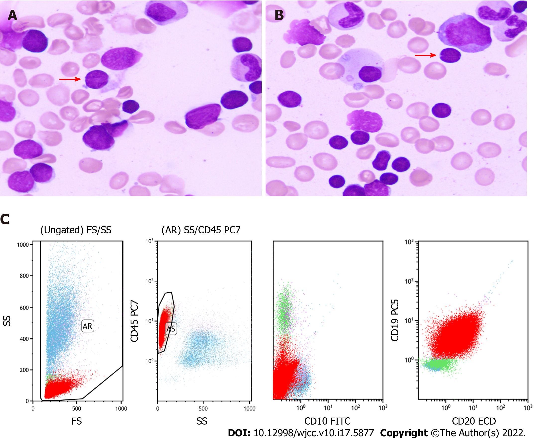
Figure 1 Morphology and flow cytometry analysis of the bone marrow.
A and B: The bone marrow (BM) was mainly infiltrated by monocytoid cells, accompanied with a few cells with cleaved or notched nuclei (pointed by arrows) [400 × (A) and 400 × (B)]; C: Flow cytometry revealed that the neoplastic cells (the red group) in BM were positive for CD19 and CD20 but negative for CD5 and CD10.
- Citation: Peng HY, Xiu YJ, Chen WH, Gu QL, Du X. Follicular lymphoma presenting like marginal zone lymphoma: A case report. World J Clin Cases 2022; 10(17): 5877-5883
- URL: https://www.wjgnet.com/2307-8960/full/v10/i17/5877.htm
- DOI: https://dx.doi.org/10.12998/wjcc.v10.i17.5877


