Copyright
©The Author(s) 2022.
World J Clin Cases. Jun 6, 2022; 10(16): 5400-5405
Published online Jun 6, 2022. doi: 10.12998/wjcc.v10.i16.5400
Published online Jun 6, 2022. doi: 10.12998/wjcc.v10.i16.5400
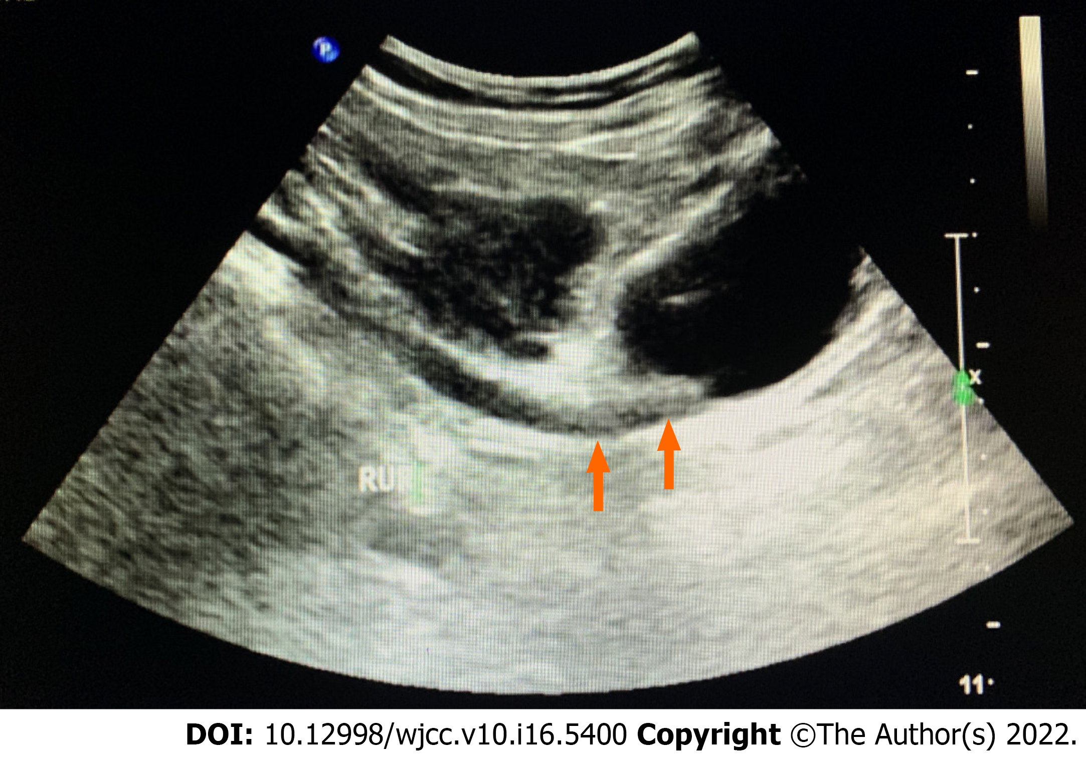
Figure 1 Orange arrows showed the right distal ureteric lesions.
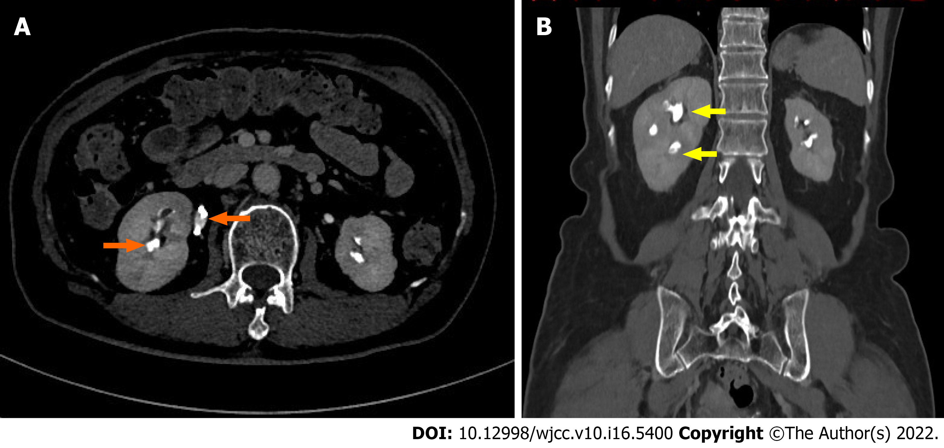
Figure 2 Computed tomography urography.
A: In axial plane, two orange arrows showed non enhancing filling defects in the upper ureter and calyces of the right kidney; B: In coronal plane, two yellow arrows showed non enhancing filling defects in the upper ureter and calyces of the right kidney.
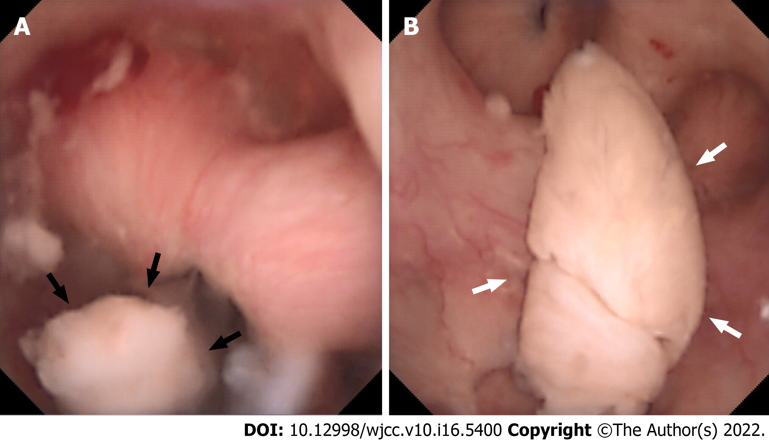
Figure 3 The endoscopic sign of renal papillary necrosis.
A: Black arrows showed the part of this renal papilla undergoing selective necrosis, which was sloughing with pedicles inside the calyces; B: White arrows showed the necrotic papilla floating as cottons in the calyces, which were characterized by whitish structures, soft and friable.
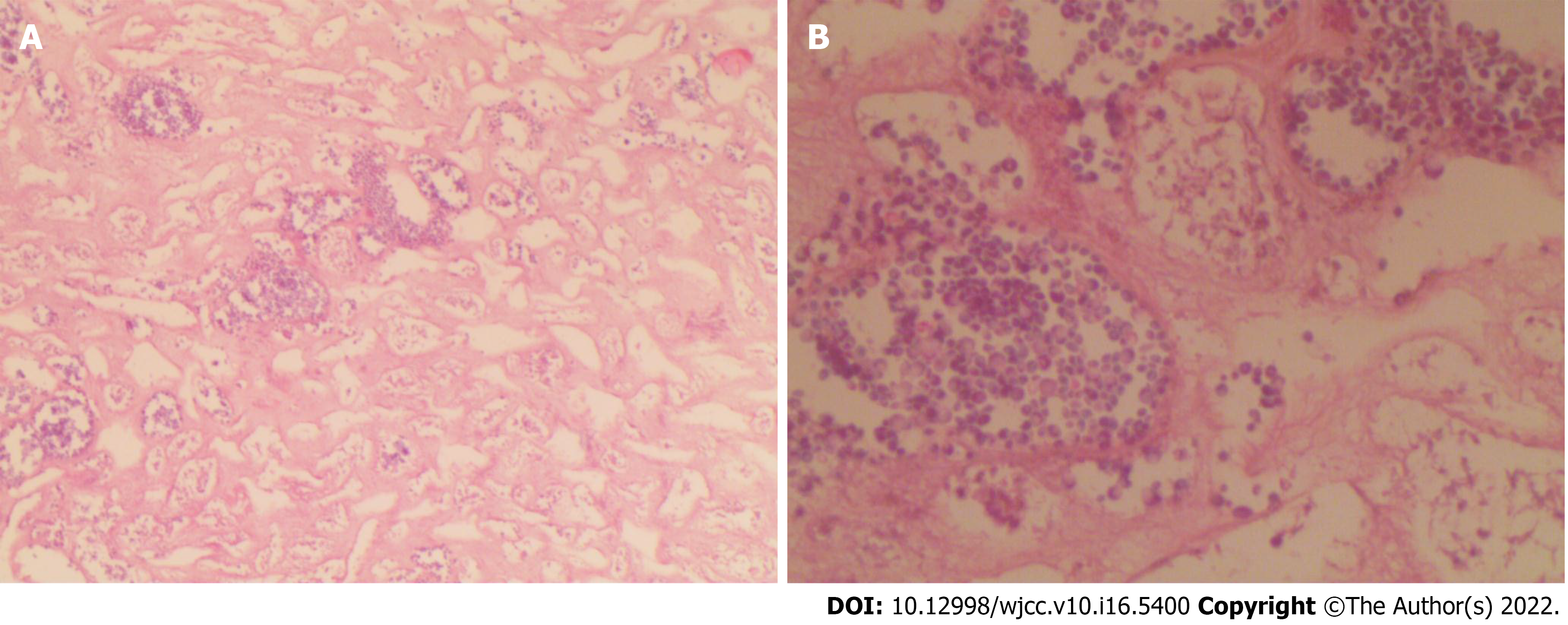
Figure 4 Histopathological findings, fragments consisting of necrotic material.
Infarcted renal papilla was accompanied with inflammatory exudations. No malignancy was identified. A: HE stain, 20 ×; B: HE stain, 40 ×.
- Citation: Pan HH, Luo YJ, Zhu QG, Ye LF. Renal papillary necrosis with urinary tract obstruction: A case report. World J Clin Cases 2022; 10(16): 5400-5405
- URL: https://www.wjgnet.com/2307-8960/full/v10/i16/5400.htm
- DOI: https://dx.doi.org/10.12998/wjcc.v10.i16.5400


