Copyright
©The Author(s) 2022.
World J Clin Cases. Jun 6, 2022; 10(16): 5400-5405
Published online Jun 6, 2022. doi: 10.12998/wjcc.v10.i16.5400
Published online Jun 6, 2022. doi: 10.12998/wjcc.v10.i16.5400
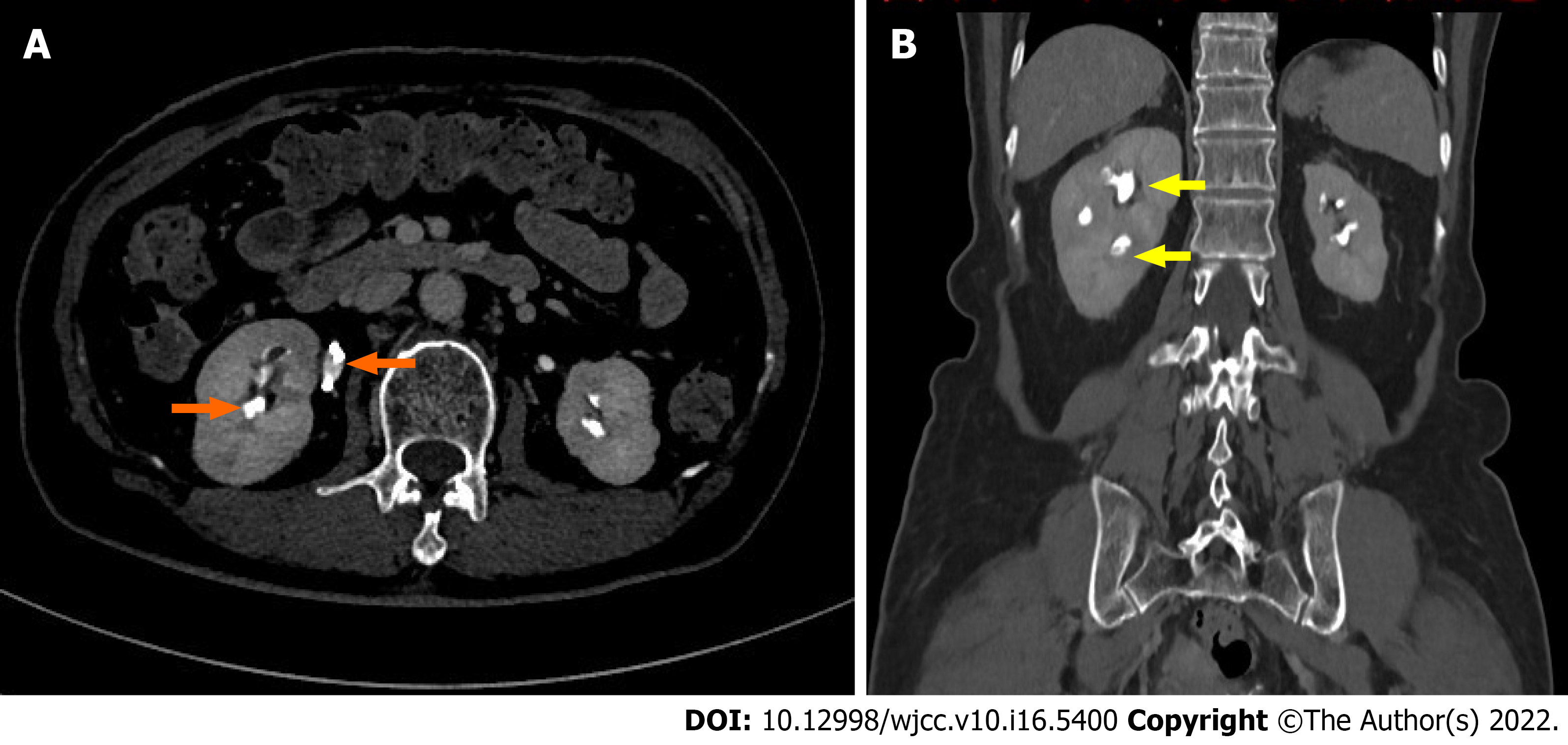
Figure 2 Computed tomography urography.
A: In axial plane, two orange arrows showed non enhancing filling defects in the upper ureter and calyces of the right kidney; B: In coronal plane, two yellow arrows showed non enhancing filling defects in the upper ureter and calyces of the right kidney.
- Citation: Pan HH, Luo YJ, Zhu QG, Ye LF. Renal papillary necrosis with urinary tract obstruction: A case report. World J Clin Cases 2022; 10(16): 5400-5405
- URL: https://www.wjgnet.com/2307-8960/full/v10/i16/5400.htm
- DOI: https://dx.doi.org/10.12998/wjcc.v10.i16.5400


