Copyright
©The Author(s) 2017.
World J Methodol. Dec 26, 2017; 7(4): 117-128
Published online Dec 26, 2017. doi: 10.5662/wjm.v7.i4.117
Published online Dec 26, 2017. doi: 10.5662/wjm.v7.i4.117
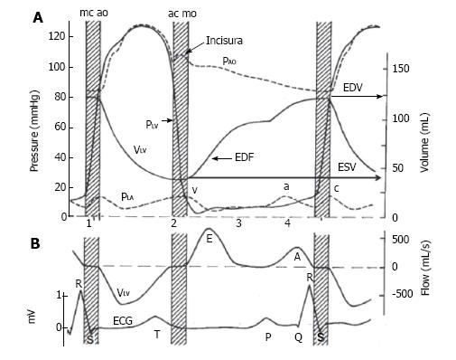
Figure 2 Describes the volume and flow relationships in the left atrium and left ventricle throughout one cardiac cycle, i.
e., systole and diastole. A: Pressure (P) and volume (V) are presented for the Aorta (Ao), left atrium (LA) and left ventricle (LV). Systole: During the early phase between mitral valve closure (mc) and aortic valve opening (ao) is the isovolumic contraction phase (stripped bar), where there is increase in PLV (solid line) without change in VLV (solid line). This is followed by ventricular contraction with a rise in PLV and PAO (upper dashed line), that peaks mid cycle, and a reduction in VLV. Diastole: At the end of LV contraction, and when the PLV is lower than the aorta the aortic valve closes (ac), followed by a period of isovolumic LV relaxation (stripped bar), where there is reduction in PLV without a change in VLV. The Incisura or dicrotic notch describes the small backflow of blood into the LV. Early diastolic point of early diastolic filling. In diastole PLA is generated early by the reservoir and conduit atrial function (v wave - lower dashed line) and corresponds with early diastolic filling (EDF) and a late atrial contraction or booster function (a wave) and contributes to late diastolic filling. Ventricular volumes are as end diastolic or end systolic (EDV or ESV; solid line). Cardiac sounds are shown as 1-4; B: Diagram showing relationship between electrical conduction and blood flow with an additional catheter in the LV. Systolic blood flow out of the ventricle (V LV), is followed by early diastolic blood flow into the LV (E wave), bate blood flow into the LV during LA contraction (A wave). A standard ECG lead II shows LA depolarization, LV depolarization, and LV repolarization (P wave, QRS complex, and T wave, respectively) (Published in Ref 28, figure provided courtesy of Dr. John V. Tyberg and Dr. Henk E. D. J. ter Keurs. Permission required). ECG: Electrocardiogram.
- Citation: Iyngkaran P, Anavekar NS, Neil C, Thomas L, Hare DL. Shortness of breath in clinical practice: A case for left atrial function and exercise stress testing for a comprehensive diastolic heart failure workup. World J Methodol 2017; 7(4): 117-128
- URL: https://www.wjgnet.com/2222-0682/full/v7/i4/117.htm
- DOI: https://dx.doi.org/10.5662/wjm.v7.i4.117


