Copyright
©2013 Baishideng Publishing Group Co.
World J Methodol. Dec 26, 2013; 3(4): 45-64
Published online Dec 26, 2013. doi: 10.5662/wjm.v3.i4.45
Published online Dec 26, 2013. doi: 10.5662/wjm.v3.i4.45
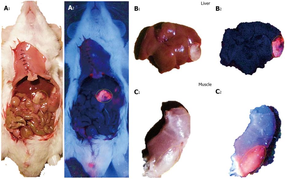
Figure 4 Macroscopic digital imaging of a mouse with acute ethanol-induced necrosis in the liver and muscle having received 5.
0 mg/kg hypericin in DMSO/PEG400/water (25:60:15, v/v/v). Under normal tungsten light, viable liver, intestines and muscle show normal appearances whereas hepatic infarction and muscle necrosis appear as white cheesy tissue (A1, B1, C1). With a UV light of 254 nm, a bright red fluorescence from liver (A2, B2) and muscle necrosis (C2) but a lack of fluorescent signal from the liver (A2, B2), viable muscle (C2) and other abdominal structures were observed (A2). DMSO: Dimethyl sulfoxide; PEG400: Polyethylene glycol 400; UV: Ultraviolet.
- Citation: Cona MM, Witte P, Verbruggen A, Ni Y. An overview of translational (radio)pharmaceutical research related to certain oncological and non-oncological applications. World J Methodol 2013; 3(4): 45-64
- URL: https://www.wjgnet.com/2222-0682/full/v3/i4/45.htm
- DOI: https://dx.doi.org/10.5662/wjm.v3.i4.45


