Copyright
©The Author(s) 2015.
World J Transl Med. Dec 12, 2015; 4(3): 88-100
Published online Dec 12, 2015. doi: 10.5528/wjtm.v4.i3.88
Published online Dec 12, 2015. doi: 10.5528/wjtm.v4.i3.88
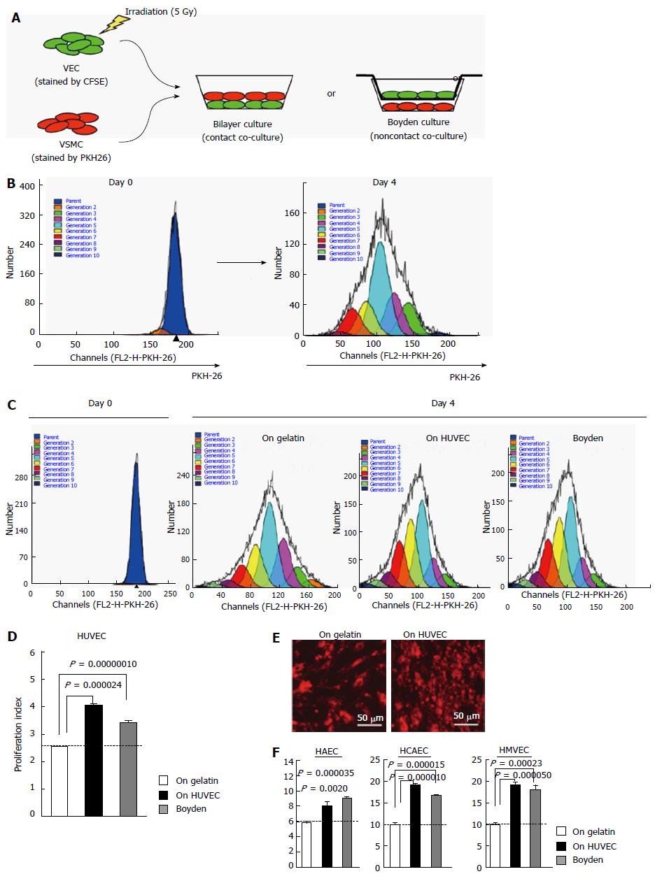
Figure 1 Phenotype determination of primary cultured human vascular endothelial cells.
A and B: Illustrations of the method to calculate proliferation indexes of VEC-co-cultured VSMC via ModFit LT™ software-based analyses; C: Results of flow cytometric analyses of HUVEC-co-cultured VSMC with ModFit LT™ analyses; D: Results in C were statistically evaluated (n = 3); E: Fluorescent microscopy of PKH-26-stained VSMC subjected to contact co-culture with HUVEC; F: Results of VSMC-co-culture experiments using HAEC, HCAEC and HMVEC (n = 3). VEC: Vascular endothelial cells; HAEC: Human umbilical artery endothelial cells; HCAEC: Human coronary artery endothelial cells; HUVEC: Human umbilical vein endothelial cells; HMVEC: Human microvascular endothelial cell; VSMC: Vascular smooth muscle cell.
- Citation: Nishio M, Nakahara M, Sato C, Saeki K, Akutsu H, Umezawa A, Tobe K, Yasuda K, Yuo A, Saeki K. New categorization of human vascular endothelial cells by pro- vs anti-proliferative phenotypes. World J Transl Med 2015; 4(3): 88-100
- URL: https://www.wjgnet.com/2220-6132/full/v4/i3/88.htm
- DOI: https://dx.doi.org/10.5528/wjtm.v4.i3.88


