Copyright
©2012 Baishideng.
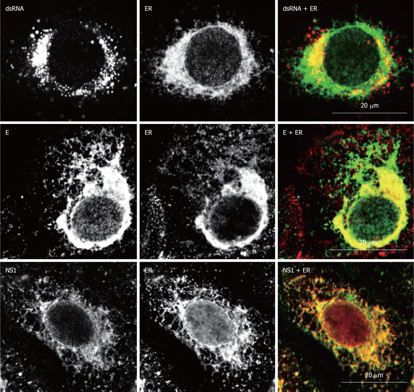
Figure 4 Localization of West Nile virus dsRNA, structural and non-structural proteins at the endoplasmic reticulum of infected cells.
Double immunofluorescence labeling of Vero cells 24h post-infection with West Nile virus (MOI of 1 PFU/cell) and stained for dsRNA, using monoclonal antibody J2 (English and Scientific Consulting Bt., Hungary), E glycoprotein or NS1, using monoclonal antibody 3.67G and 3.1112G (Millipore, Temecula, CA), respectively, in combination with rabbit polyclonal antibody against calnexin (Abcam, Cambridge UK) as an specific marker for endoplasmic reticulum. Procedures for immunostaining have been previously described[21].
- Citation: Martín-Acebes MA, Saiz JC. West Nile virus: A re-emerging pathogen revisited. World J Virol 2012; 1(2): 51-70
- URL: https://www.wjgnet.com/2220-3249/full/v1/i2/51.htm
- DOI: https://dx.doi.org/10.5501/wjv.v1.i2.51


