Copyright
©The Author(s) 2025.
World J Psychiatry. Jun 19, 2025; 15(6): 105751
Published online Jun 19, 2025. doi: 10.5498/wjp.v15.i6.105751
Published online Jun 19, 2025. doi: 10.5498/wjp.v15.i6.105751
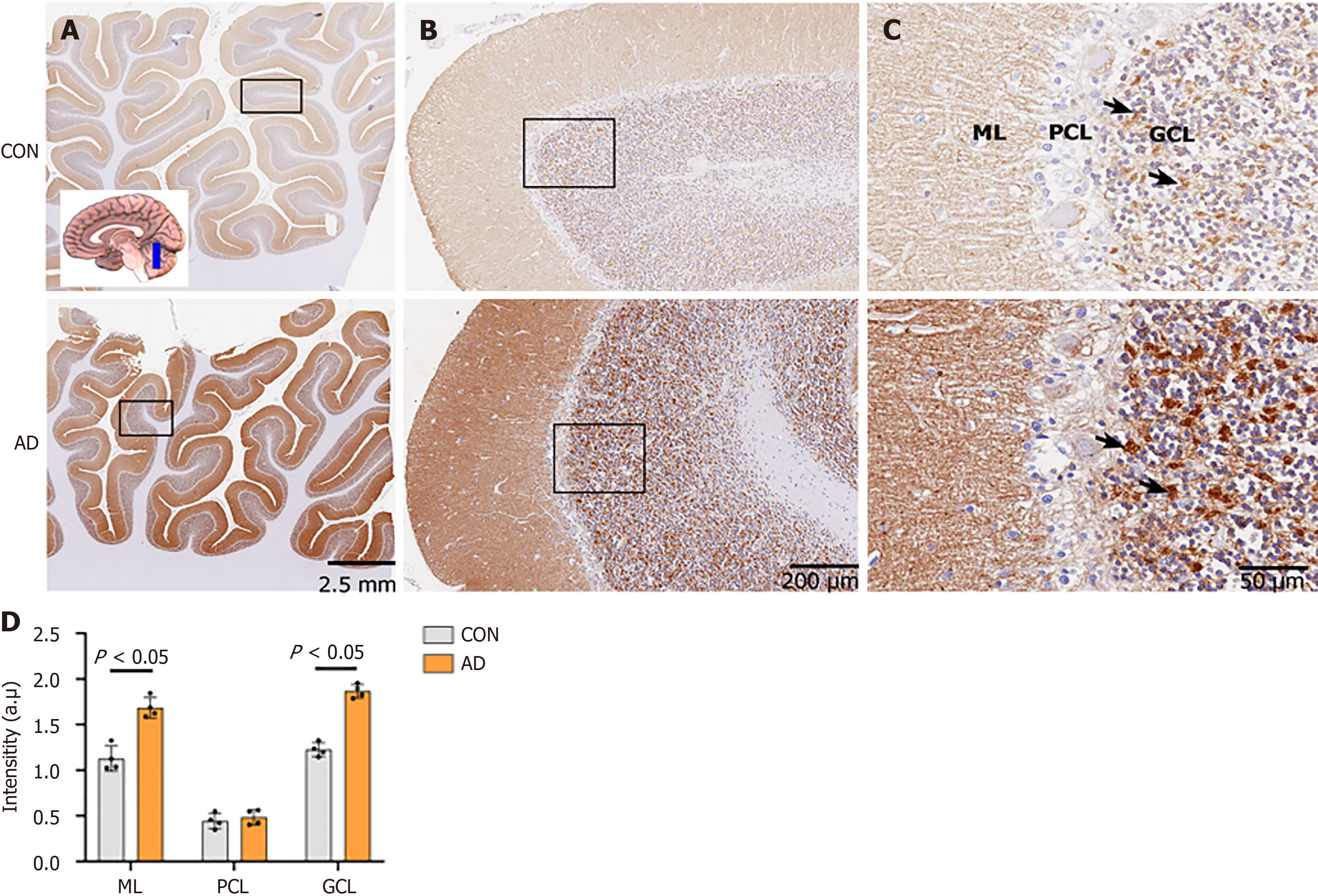
Figure 4 Neuroplastin 65 immunoreactive pattern in the cerebellum derived from individuals with Alzheimer’s disease and age-matched controls.
A: Low-power microphotograph showing the regional distribution of neuroplastin 65 (NP65) immunoreactivity in the cerebellar anterior lobe of controls (CON) and Alzheimer's disease (AD). The inset indicates the position of the sampling (scale bars = 2.5 mm); B: Low-power views in the rectangle frame in Figure 4A showing the regional distribution of NP65 in the cerebellar cortex of CON and AD (scale bars = 200 μm); C: Higher-power views in the ML, PCL and GCL of the rectangle frame in Figure 4B. Note that intense NP65 immunoreactivity is shown in a blocky or granular pattern (indicated by arrows) in the GCL, while it is punctate in the ML. There is a significant increase in immunoreactivity in AD compared with CON. The nucleus is stained with hematoxylin (scale bars = 50 μm); D: Quantification of NP65 immunoreactivity in the cerebellar cortex between AD and CON. CON: Controls; AD: Alzheimer's disease; GCL: Granular cell layer; PCL: Purkinje cell layer; ML: Molecular layer.
- Citation: Zheng YN, Wang Y, Chen L, Xu LZ, Zhang L, Wang JL, Liu J, Zhang QL, Yuan QL. Increased expression of the neuroplastin 65 protein is involved in neurofibrillary tangles and amyloid beta plaques in Alzheimer’s disease. World J Psychiatry 2025; 15(6): 105751
- URL: https://www.wjgnet.com/2220-3206/full/v15/i6/105751.htm
- DOI: https://dx.doi.org/10.5498/wjp.v15.i6.105751


