Copyright
©The Author(s) 2016.
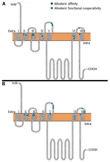
Figure 2 Snake diagram of muscarinic M1 receptor.
The snake diagrams begin with the extracellular amino terminal and terminate at the intracellular carboxyl terminal. Highlighted are amino acids identified by site directed mutagenesis as being implicated in (A) allosteric ligand binding (green) and (B) functional cooperativity (blue)[94-96]. Roman numerals denote transmembrane domains. Diagrams generated using Protter[113]. Extra: Extracellular domain; Intra: Intracellular domain.
- Citation: Hopper S, Udawela M, Scarr E, Dean B. Allosteric modulation of cholinergic system: Potential approach to treating cognitive deficits of schizophrenia. World J Pharmacol 2016; 5(1): 32-43
- URL: https://www.wjgnet.com/2220-3192/full/v5/i1/32.htm
- DOI: https://dx.doi.org/10.5497/wjp.v5.i1.32


