Copyright
©2013 Baishideng Publishing Group Co.
World J Surg Proced. Nov 28, 2013; 3(3): 54-59
Published online Nov 28, 2013. doi: 10.5412/wjsp.v3.i3.54
Published online Nov 28, 2013. doi: 10.5412/wjsp.v3.i3.54
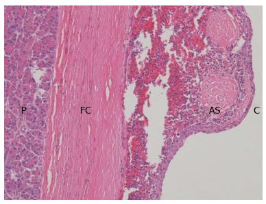
Figure 4 Microscopically, the cyst (C) was covered with stratified squamous epithelium and was surrounded by normal splenic tissue.
A fibrous capsule (FC) separates the intrapancreatic accessory spleen (AS) from pancreas (P) (HE, × 100).
- Citation: Lee CL, Di Y, Jiang YJ, Jin C, Fu DL. Epidermoid cyst of intrapancreatic accessory spleen: A case report and literature review. World J Surg Proced 2013; 3(3): 54-59
- URL: https://www.wjgnet.com/2219-2832/full/v3/i3/54.htm
- DOI: https://dx.doi.org/10.5412/wjsp.v3.i3.54


