
















©The Author(s) 2017.
World J Clin Pediatr. Feb 8, 2017; 6(1): 10-23
Published online Feb 8, 2017. doi: 10.5409/wjcp.v6.i1.10
Published online Feb 8, 2017. doi: 10.5409/wjcp.v6.i1.10
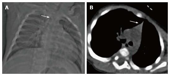
Figure 8 Thymic hypoplasia in a 3-mo-old male infant with primary immunodeficiency.
CT scanogram (A) reveals small size of the thymus, i.e., less than twice the width of the third thoracic vertebra (arrow). NCCT chest axial section (B) shows triangular configuration of the thymus (arrow) with straight margins (normal appearance in adolescents). Compare this with the normal appearance of the thymus in a 2-mo-old infant (Figure 6A and B).
- Citation: Manchanda S, Bhalla AS, Jana M, Gupta AK. Imaging of the pediatric thymus: Clinicoradiologic approach. World J Clin Pediatr 2017; 6(1): 10-23
- URL: https://www.wjgnet.com/2219-2808/full/v6/i1/10.htm
- DOI: https://dx.doi.org/10.5409/wjcp.v6.i1.10