Copyright
©The Author(s) 2016.
World J Clin Pediatr. May 8, 2016; 5(2): 228-233
Published online May 8, 2016. doi: 10.5409/wjcp.v5.i2.228
Published online May 8, 2016. doi: 10.5409/wjcp.v5.i2.228
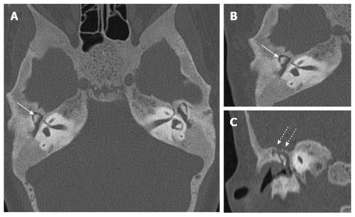
Figure 2 Axial high resolution computed tomography images.
(A and B) showing soft tissue opacification in bilateral middle ears (A) with erosion of head of head of malleus (arrows). Coronal reformatted CT image (C) showing thinning and erosion of tegmen tympani (dotted arrows) secondary to cholesteatoma.
- Citation: Verma R, Jana M, Bhalla AS, Kumar A, Kumar R. Diagnosis of osteopetrosis in bilateral congenital aural atresia: Turning point in treatment strategy. World J Clin Pediatr 2016; 5(2): 228-233
- URL: https://www.wjgnet.com/2219-2808/full/v5/i2/228.htm
- DOI: https://dx.doi.org/10.5409/wjcp.v5.i2.228


