Copyright
©The Author(s) 2016.
World J Clin Pediatr. Feb 8, 2016; 5(1): 1-15
Published online Feb 8, 2016. doi: 10.5409/wjcp.v5.i1.1
Published online Feb 8, 2016. doi: 10.5409/wjcp.v5.i1.1
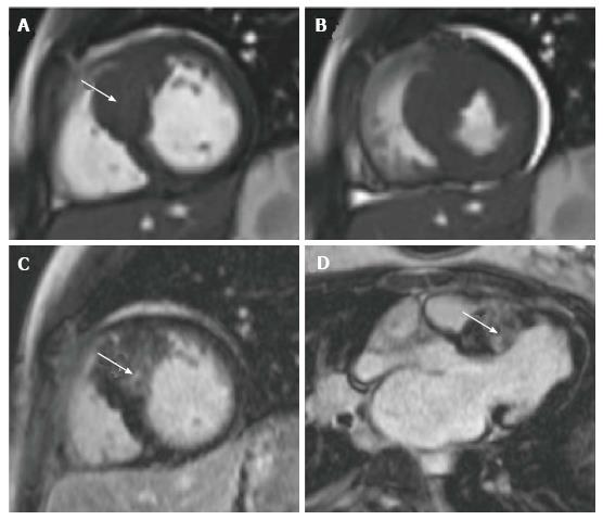
Figure 4 A new diagnosis of hypertrophic cardiomyopathy in a 12-year-old girl who presented as an out of hospital arrest with documented ventricular fibrillation.
A shows a short axis view of the left ventricle at end systole at basal level. There is asymmetrical septal hypertrophy (arrowed) with a maximal wall thickness of 26 mm; B shows the same ventricular position in end systole; C and D are late gadolinium enhancement images demonstrating extensive patchy fibrosis of the hypertrophied septum (arrowed).
- Citation: Mitchell FM, Prasad SK, Greil GF, Drivas P, Vassiliou VS, Raphael CE. Cardiovascular magnetic resonance: Diagnostic utility and specific considerations in the pediatric population. World J Clin Pediatr 2016; 5(1): 1-15
- URL: https://www.wjgnet.com/2219-2808/full/v5/i1/1.htm
- DOI: https://dx.doi.org/10.5409/wjcp.v5.i1.1


