Copyright
©The Author(s) 2016.
World J Clin Pediatr. Feb 8, 2016; 5(1): 1-15
Published online Feb 8, 2016. doi: 10.5409/wjcp.v5.i1.1
Published online Feb 8, 2016. doi: 10.5409/wjcp.v5.i1.1
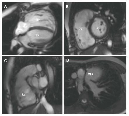
Figure 3 A 10-year-old girl with tetralogy of Fallot repaired at 1 year of age.
She had a 1 year history of increasing exertional breathlessness and reduced exercise capacity. Her CMR showed near-free pulmonary regurgitation with a dilated right ventricle and a reduced right ventricular ejection fraction. In view of these findings, she was offered a pulmonary valve replacement. A shows a 4 chamber SSFP cine image; B a short axis through the mid ventricle; C a right ventricular outflow tract view and D a transverse section through the thorax at main pulmonary artery level showing the dilated main pulmonary artery. RV: Right ventricle; LV: Left ventricle; MPA: Main pulmonary artery; SSFP: Steady state free precession; CMR: Cardiovascular magnetic resonance.
- Citation: Mitchell FM, Prasad SK, Greil GF, Drivas P, Vassiliou VS, Raphael CE. Cardiovascular magnetic resonance: Diagnostic utility and specific considerations in the pediatric population. World J Clin Pediatr 2016; 5(1): 1-15
- URL: https://www.wjgnet.com/2219-2808/full/v5/i1/1.htm
- DOI: https://dx.doi.org/10.5409/wjcp.v5.i1.1


