
















©The Author(s) 2016.
World J Clin Pediatr. Feb 8, 2016; 5(1): 1-15
Published online Feb 8, 2016. doi: 10.5409/wjcp.v5.i1.1
Published online Feb 8, 2016. doi: 10.5409/wjcp.v5.i1.1
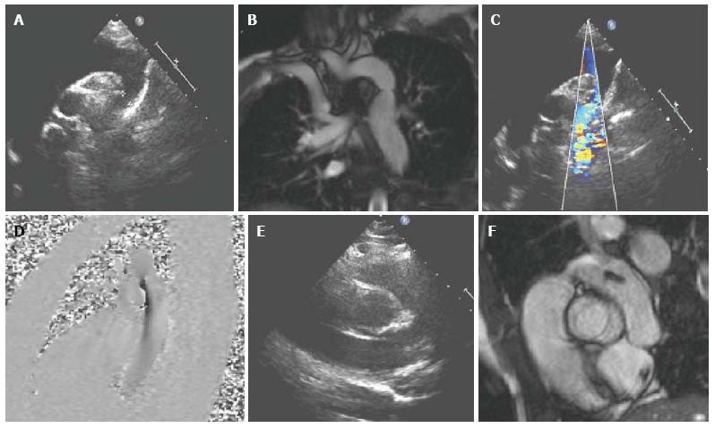
Figure 2 A 17-year-old girl with coarctation of the descending aorta, imaged by transthoracic echocardiography (A,C,E) and cardiovascular magnetic resonance (B,D,F).
Imaging of the aortic arch using TTE can be challenging, depending on the suprasternal image quality A and especially in older children with poorer acoustic windows. CMR allows accurate measurement of the dimensions of the area of stenosis B and assessment of the remainder of the thoracic aorta and head and neck vessels. Flow velocity mapping D, similar to Doppler echocardiography C is used to measure peak velocity through the stenosed area and assess for a diastolic tail. In this case, the bicuspid aortic valve was not well visualised on TTE E but was clearly seen on CMR F. CMR: Cardiovascular magnetic resonance; TTE: Transthoracic echocardiography.
- Citation: Mitchell FM, Prasad SK, Greil GF, Drivas P, Vassiliou VS, Raphael CE. Cardiovascular magnetic resonance: Diagnostic utility and specific considerations in the pediatric population. World J Clin Pediatr 2016; 5(1): 1-15
- URL: https://www.wjgnet.com/2219-2808/full/v5/i1/1.htm
- DOI: https://dx.doi.org/10.5409/wjcp.v5.i1.1