
















Copyright: ©Author(s) 2026.
World J Clin Pediatr. Jun 9, 2026; 15(2): 113603
Published online Jun 9, 2026. doi: 10.5409/wjcp.v15.i2.113603
Published online Jun 9, 2026. doi: 10.5409/wjcp.v15.i2.113603
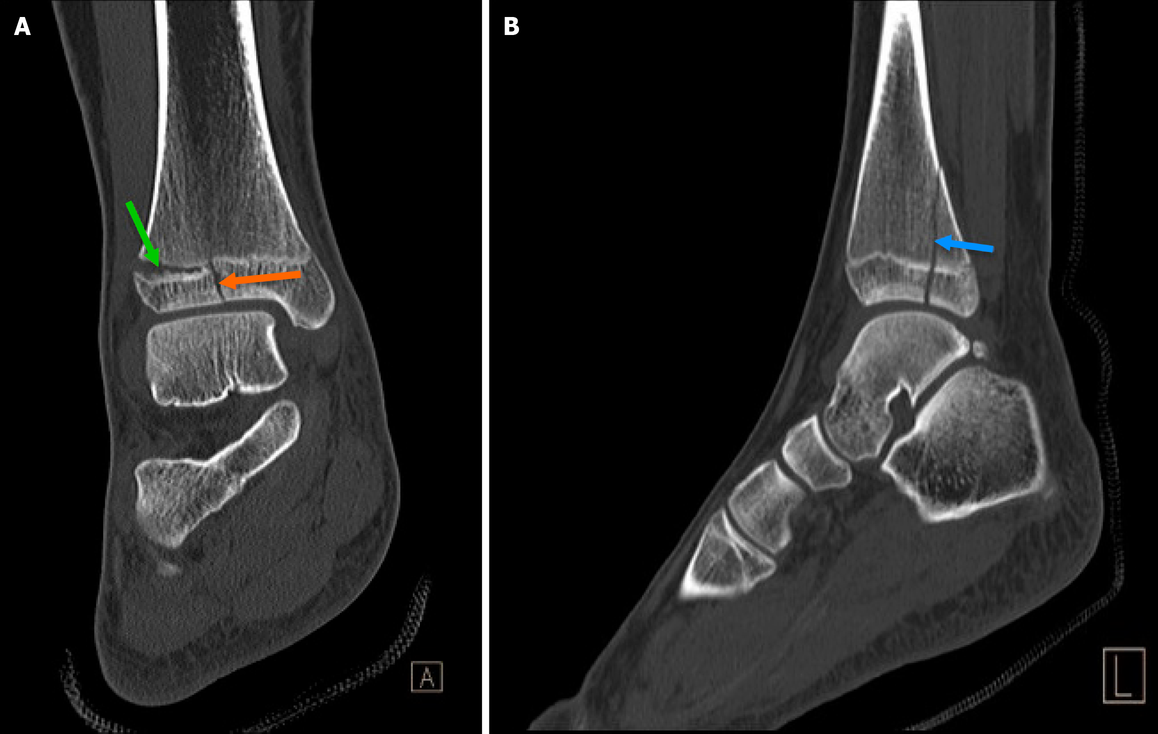
Figure 4 This is a triplane fracture (fracture across 3 planes).
A: Sagittal (orange arrow), axial (green arrow); B: Coronal (blue arrow) fracture. The coronal fracture can be seen on the sagittal view in the image on the right[12]. Citation: Al Salam H. Triplanar fracture. Case study, Radiopaedia.org 2015. Copyright ©The Authors 2015. Published by Radiopaedia. The authors have obtained the permission (Supplementary material).
- Citation: Perera Molligoda Arachchige AS, Schmiliver B, Patel HA. Imaging of pediatric musculoskeletal trauma: Age-specific considerations and challenges. World J Clin Pediatr 2026; 15(2): 113603
- URL: https://www.wjgnet.com/2219-2808/full/v15/i2/113603.htm
- DOI: https://dx.doi.org/10.5409/wjcp.v15.i2.113603