Copyright
©The Author(s) 2025.
World J Clin Pediatr. Sep 9, 2025; 14(3): 103732
Published online Sep 9, 2025. doi: 10.5409/wjcp.v14.i3.103732
Published online Sep 9, 2025. doi: 10.5409/wjcp.v14.i3.103732
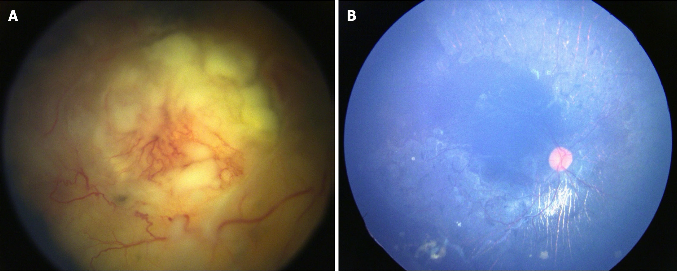
Figure 1 Regression pattern.
A: Fundus image of the right eye showed group D retinoblastoma with diffuse seeds with optic disc obscuration at presentation; B: After one cycle of primary intra-arterial chemotherapy, the calcified tumor near the inferior arcade with chorioretinal atrophic changes and optic disc were visible.
- Citation: Das A, Saiteja K, Shah PK, Prema S, Narendran V. Outcomes and adverse events following intra-arterial chemotherapy for retinoblastoma: A single center study in South India. World J Clin Pediatr 2025; 14(3): 103732
- URL: https://www.wjgnet.com/2219-2808/full/v14/i3/103732.htm
- DOI: https://dx.doi.org/10.5409/wjcp.v14.i3.103732


