
















©The Author(s) 2022.
World J Clin Pediatr. Jan 9, 2022; 11(1): 71-84
Published online Jan 9, 2022. doi: 10.5409/wjcp.v11.i1.71
Published online Jan 9, 2022. doi: 10.5409/wjcp.v11.i1.71
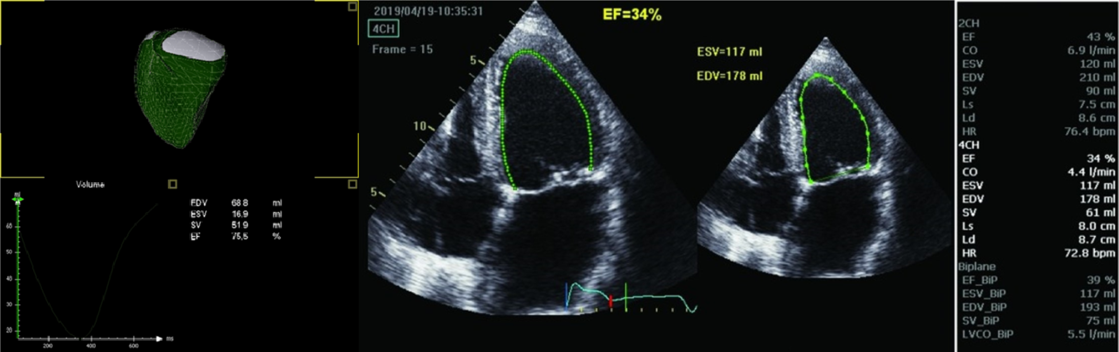
Figure 1 Three-dimensional echocardiographic reconstruction of the delineation of right ventricular ejection fraction.
The endocardial border is traced throughout the cardiac cycle using speckle tracking, and the software automatically locates end-systolic and end-diastolic frames. Example of a four-dimensional echocardiographic reconstruction of the delineation of the right ventricle seen from the septal side (end-diastolic volume 68.8 mL, end-systolic volume 16.9 mL, and ejection fraction 75.5%). The mesh is the right ventricle at end-diastole, in green at end-systole. The pulmonary valve is shown in white on the upper left side, the tricuspid valve is shown on the upper right side, and the right ventricular apex toward the bottom.
- Citation: Al-Biltagi M, Elrazaky O, Mawlana W, Srour E, Shabana AH. Tissue Doppler, speckling tracking and four-dimensional echocardiographic assessment of right ventricular function in children with dilated cardiomyopathy. World J Clin Pediatr 2022; 11(1): 71-84
- URL: https://www.wjgnet.com/2219-2808/full/v11/i1/71.htm
- DOI: https://dx.doi.org/10.5409/wjcp.v11.i1.71